• 文献检索
  • 文档翻译
  • 深度研究
  • 学术资讯
  • Suppr Zotero 插件Zotero 插件
  • 邀请有礼
  • 套餐&价格
  • 历史记录
应用&插件
Suppr Zotero 插件Zotero 插件浏览器插件Mac 客户端Windows 客户端微信小程序
定价
高级版会员购买积分包购买API积分包
服务
文献检索文档翻译深度研究API 文档MCP 服务
关于我们
关于 Suppr公司介绍联系我们用户协议隐私条款
关注我们

Suppr 超能文献

核心技术专利:CN118964589B侵权必究
粤ICP备2023148730 号-1Suppr @ 2026

文献检索

告别复杂PubMed语法,用中文像聊天一样搜索,搜遍4000万医学文献。AI智能推荐,让科研检索更轻松。

立即免费搜索

文件翻译

保留排版,准确专业,支持PDF/Word/PPT等文件格式,支持 12+语言互译。

免费翻译文档

深度研究

AI帮你快速写综述,25分钟生成高质量综述,智能提取关键信息,辅助科研写作。

立即免费体验

利用新型 CT 序列选择算法优化卵巢肿瘤分类。

Optimising ovarian tumor classification using a novel CT sequence selection algorithm.

机构信息

Department of Information Science & Engineering, Bapuji Institute of Engineering & Technology, Davanagere, Karnataka, India.

Department of Information Technology, Faculty of Computing and Information Technology, King Abdulaziz University, Jeddah, Saudi Arabia.

出版信息

Sci Rep. 2024 Oct 23;14(1):25010. doi: 10.1038/s41598-024-75555-2.

DOI:10.1038/s41598-024-75555-2
PMID:39443517
原文链接:https://pmc.ncbi.nlm.nih.gov/articles/PMC11500337/
Abstract

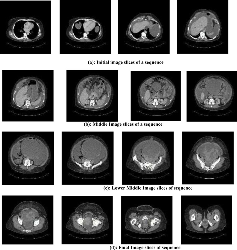
Gynaecological cancers, especially ovarian cancer, remain a critical public health issue, particularly in regions like India, where there are challenges related to cancer awareness, variable pathology, and limited access to screening facilities. These challenges often lead to the diagnosis of cancer at advanced stages, resulting in poorer outcomes for patients. The goal of this study is to enhance the accuracy of classifying ovarian tumours, with a focus on distinguishing between malignant and early-stage cases, by applying advanced deep learning methods. In our approach, we utilized three pre-trained deep learning models-Xception, ResNet50V2, and ResNet50V2FPN-to classify ovarian tumors using publicly available Computed Tomography (CT) scan data. To further improve the model's performance, we developed a novel CT Sequence Selection Algorithm, which optimises the use of CT images for a more precise classification of ovarian tumours. The models were trained and evaluated on selected TIFF images, comparing the performance of the ResNet50V2FPN model with and without the CT Sequence Selection Algorithm. Our experimental results show the Comparative evaluation against the ResNet50V2 FPN model, both with and without the CT Sequence Selection Algorithm, demonstrates the superiority of the proposed algorithm over existing state-of-the-art methods. This research presents a promising approach for improving the early detection and management of gynecological cancers, with potential benefits for patient outcomes, especially in areas with limited healthcare resources.

摘要

妇科癌症,尤其是卵巢癌,仍然是一个严重的公共卫生问题,特别是在印度等地区,这些地区在癌症意识、多变的病理学和有限的筛查设施方面存在挑战。这些挑战往往导致癌症在晚期被诊断出来,从而导致患者的预后更差。本研究的目的是通过应用先进的深度学习方法来提高卵巢肿瘤分类的准确性,特别是区分恶性和早期病例。在我们的方法中,我们使用了三个预先训练的深度学习模型——Xception、ResNet50V2 和 ResNet50V2FPN,使用公开的计算机断层扫描 (CT) 扫描数据来对卵巢肿瘤进行分类。为了进一步提高模型的性能,我们开发了一种新的 CT 序列选择算法,该算法优化了 CT 图像的使用,以更精确地对卵巢肿瘤进行分类。我们在选定的 TIFF 图像上对模型进行了训练和评估,比较了使用和不使用 CT 序列选择算法的 ResNet50V2FPN 模型的性能。我们的实验结果表明,与 ResNet50V2 FPN 模型相比,无论是使用还是不使用 CT 序列选择算法,该算法的性能都优于现有的最先进方法。这项研究提出了一种有前途的方法,可以改善妇科癌症的早期检测和管理,特别是在医疗资源有限的地区,对患者的预后有潜在的好处。

https://cdn.ncbi.nlm.nih.gov/pmc/blobs/71fd/11500337/933bc36d580a/41598_2024_75555_Fig15_HTML.jpg
https://cdn.ncbi.nlm.nih.gov/pmc/blobs/71fd/11500337/59e941b77b90/41598_2024_75555_Fig1_HTML.jpg
https://cdn.ncbi.nlm.nih.gov/pmc/blobs/71fd/11500337/3f62dd98cd46/41598_2024_75555_Fig2_HTML.jpg
https://cdn.ncbi.nlm.nih.gov/pmc/blobs/71fd/11500337/5aca85af2af5/41598_2024_75555_Fig3_HTML.jpg
https://cdn.ncbi.nlm.nih.gov/pmc/blobs/71fd/11500337/a1eb049208cc/41598_2024_75555_Fig4_HTML.jpg
https://cdn.ncbi.nlm.nih.gov/pmc/blobs/71fd/11500337/5f11e212c821/41598_2024_75555_Fig5_HTML.jpg
https://cdn.ncbi.nlm.nih.gov/pmc/blobs/71fd/11500337/d32d522df4ec/41598_2024_75555_Fig6_HTML.jpg
https://cdn.ncbi.nlm.nih.gov/pmc/blobs/71fd/11500337/9a98da37decd/41598_2024_75555_Fig7_HTML.jpg
https://cdn.ncbi.nlm.nih.gov/pmc/blobs/71fd/11500337/a3152389fa13/41598_2024_75555_Fig8_HTML.jpg
https://cdn.ncbi.nlm.nih.gov/pmc/blobs/71fd/11500337/dab3d1c0b130/41598_2024_75555_Fig9_HTML.jpg
https://cdn.ncbi.nlm.nih.gov/pmc/blobs/71fd/11500337/3fe3c0f0d0c2/41598_2024_75555_Fig10_HTML.jpg
https://cdn.ncbi.nlm.nih.gov/pmc/blobs/71fd/11500337/2ddb58d611a6/41598_2024_75555_Fig11_HTML.jpg
https://cdn.ncbi.nlm.nih.gov/pmc/blobs/71fd/11500337/6480699c7f33/41598_2024_75555_Fig12_HTML.jpg
https://cdn.ncbi.nlm.nih.gov/pmc/blobs/71fd/11500337/3e5e45694923/41598_2024_75555_Fig13_HTML.jpg
https://cdn.ncbi.nlm.nih.gov/pmc/blobs/71fd/11500337/d0b65ffe6148/41598_2024_75555_Figa_HTML.jpg
https://cdn.ncbi.nlm.nih.gov/pmc/blobs/71fd/11500337/bf4b70654025/41598_2024_75555_Fig14_HTML.jpg
https://cdn.ncbi.nlm.nih.gov/pmc/blobs/71fd/11500337/933bc36d580a/41598_2024_75555_Fig15_HTML.jpg

相似文献

1
Optimising ovarian tumor classification using a novel CT sequence selection algorithm.利用新型 CT 序列选择算法优化卵巢肿瘤分类。
Sci Rep. 2024 Oct 23;14(1):25010. doi: 10.1038/s41598-024-75555-2.
2
Enhancing Alzheimer's Disease Classification with Transfer Learning: Finetuning a Pre-trained Algorithm.利用迁移学习增强阿尔茨海默病分类:微调预训练算法。
Curr Med Imaging. 2024;20:e15734056305633. doi: 10.2174/0115734056305633240603061644.
3
An automated diagnosis and classification of COVID-19 from chest CT images using a transfer learning-based convolutional neural network.利用基于迁移学习的卷积神经网络对 chest CT 图像进行 COVID-19 的自动诊断和分类。
Comput Biol Med. 2022 May;144:105383. doi: 10.1016/j.compbiomed.2022.105383. Epub 2022 Mar 10.
4
Ultrasound-based deep learning radiomics model for differentiating benign, borderline, and malignant ovarian tumours: a multi-class classification exploratory study.基于超声的深度学习放射组学模型用于鉴别良性、交界性和恶性卵巢肿瘤:多分类分类探索性研究。
BMC Med Imaging. 2024 Apr 15;24(1):89. doi: 10.1186/s12880-024-01251-2.
5
Automatic Detection and Segmentation of Ovarian Cancer Using a Multitask Model in Pelvic CT Images.基于盆腔 CT 图像的多任务模型自动检测和分割卵巢癌。
Oxid Med Cell Longev. 2022 Oct 11;2022:6009107. doi: 10.1155/2022/6009107. eCollection 2022.
6
Fully automatic deep learning-based lung parenchyma segmentation and boundary correction in thoracic CT scans.基于深度学习的全自动化肺部组织分割和胸 CT 扫描边界修正。
Int J Comput Assist Radiol Surg. 2024 Feb;19(2):261-272. doi: 10.1007/s11548-023-03010-0. Epub 2023 Aug 18.
7
Automatic detection and classification of lung cancer CT scans based on deep learning and ebola optimization search algorithm.基于深度学习和埃博拉优化搜索算法的肺癌 CT 扫描自动检测和分类。
PLoS One. 2023 Aug 17;18(8):e0285796. doi: 10.1371/journal.pone.0285796. eCollection 2023.
8
Pattern Classification for Ovarian Tumors by Integration of Radiomics and Deep Learning Features.通过整合影像组学和深度学习特征对卵巢肿瘤进行模式分类
Curr Med Imaging. 2022;18(14):1486-1502. doi: 10.2174/1573405618666220516122145.
9
A deep learning- and partial least square regression-based model observer for a low-contrast lesion detection task in CT.基于深度学习和偏最小二乘回归的 CT 低对比度病灶检测任务模型观察器。
Med Phys. 2019 May;46(5):2052-2063. doi: 10.1002/mp.13500. Epub 2019 Apr 1.
10
Application of deep learning to the diagnosis of cervical lymph node metastasis from thyroid cancer with CT: external validation and clinical utility for resident training.深度学习在 CT 诊断甲状腺癌颈淋巴结转移中的应用:外部验证和对住院医师培训的临床实用性。
Eur Radiol. 2020 Jun;30(6):3066-3072. doi: 10.1007/s00330-019-06652-4. Epub 2020 Feb 17.

引用本文的文献

1
A pioneering artificial intelligence tool to predict treatment outcomes in ovarian cancer via diagnostic laparoscopy.一种通过诊断性腹腔镜检查预测卵巢癌治疗结果的开创性人工智能工具。
Sci Rep. 2025 Apr 25;15(1):14437. doi: 10.1038/s41598-025-98434-w.
2
Multiple perception contrastive learning for automated ovarian tumor classification in CT images.用于CT图像中卵巢肿瘤自动分类的多感知对比学习
Abdom Radiol (NY). 2025 Mar 12. doi: 10.1007/s00261-025-04879-y.

本文引用的文献

1
A hybrid cancer prediction based on multi-omics data and reinforcement learning state action reward state action (SARSA).基于多组学数据和强化学习状态动作奖励状态动作 (SARSA) 的混合癌症预测。
Comput Biol Med. 2023 Mar;154:106617. doi: 10.1016/j.compbiomed.2023.106617. Epub 2023 Feb 3.
2
Assessment of Sexual Dysfunction in Cervical Cancer Patients after Different Treatment Modality: A Systematic Review.评估不同治疗方式对宫颈癌患者性功能障碍的影响:系统评价。
Medicina (Kaunas). 2022 Sep 5;58(9):1223. doi: 10.3390/medicina58091223.
3
Attentional feature pyramid network for small object detection.
注意特征金字塔网络用于小目标检测。
Neural Netw. 2022 Nov;155:439-450. doi: 10.1016/j.neunet.2022.08.029. Epub 2022 Sep 5.
4
Patterns of Recurrent Disease in Cervical Cancer.宫颈癌复发疾病模式
J Pers Med. 2022 May 6;12(5):755. doi: 10.3390/jpm12050755.
5
Breast Cancer Classification from Ultrasound Images Using Probability-Based Optimal Deep Learning Feature Fusion.基于概率最优深度学习特征融合的超声图像乳腺癌分类
Sensors (Basel). 2022 Jan 21;22(3):807. doi: 10.3390/s22030807.
6
Molecular imaging in oncology: Current impact and future directions.肿瘤学中的分子影像学:当前影响和未来方向。
CA Cancer J Clin. 2022 Jul;72(4):333-352. doi: 10.3322/caac.21713. Epub 2021 Dec 13.
7
Preoperative staging of ovarian cancer: comparison between ultrasound, CT and whole-body diffusion-weighted MRI (ISAAC study).卵巢癌的术前分期:超声、CT与全身扩散加权磁共振成像的比较(ISAAC研究)
Ultrasound Obstet Gynecol. 2022 Feb;59(2):248-262. doi: 10.1002/uog.23654.
8
Multimodality Imaging Approach to Ovarian Neoplasms with Pathologic Correlation.多模态影像学方法与卵巢肿瘤的病理相关性。
Radiographics. 2021 Jan-Feb;41(1):289-315. doi: 10.1148/rg.2021200086. Epub 2020 Nov 13.
9
A modified deep convolutional neural network for detecting COVID-19 and pneumonia from chest X-ray images based on the concatenation of Xception and ResNet50V2.一种基于Xception和ResNet50V2拼接的用于从胸部X光图像中检测新冠肺炎和肺炎的改进型深度卷积神经网络。
Inform Med Unlocked. 2020;19:100360. doi: 10.1016/j.imu.2020.100360. Epub 2020 May 26.
10
Feature enhancement framework for brain tumor segmentation and classification.用于脑肿瘤分割与分类的特征增强框架。
Microsc Res Tech. 2019 Jun;82(6):803-811. doi: 10.1002/jemt.23224. Epub 2019 Feb 15.