Baker Gregory J, Novikov Edward, Zhao Ziyuan, Vallius Tuulia, Davis Janae A, Lin Jia-Ren, Muhlich Jeremy L, Mittendorf Elizabeth A, Santagata Sandro, Guerriero Jennifer L, Sorger Peter K
Ludwig Center for Cancer Research at Harvard, Harvard Medical School, Boston, MA, USA.
Laboratory of Systems Pharmacology, Program in Therapeutic Science, Harvard Medical School, Boston, MA, USA.
Nat Methods. 2024 Dec;21(12):2248-2259. doi: 10.1038/s41592-024-02328-0. Epub 2024 Oct 30.
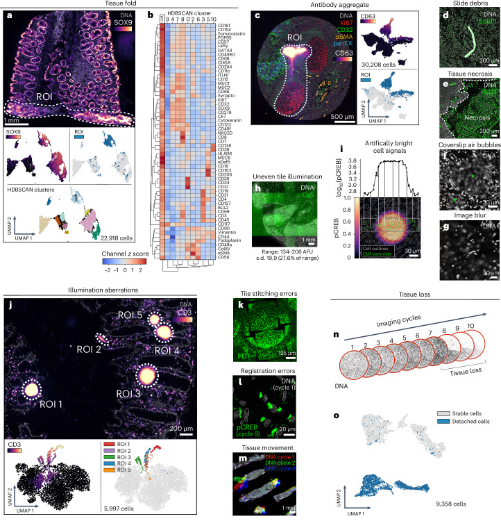
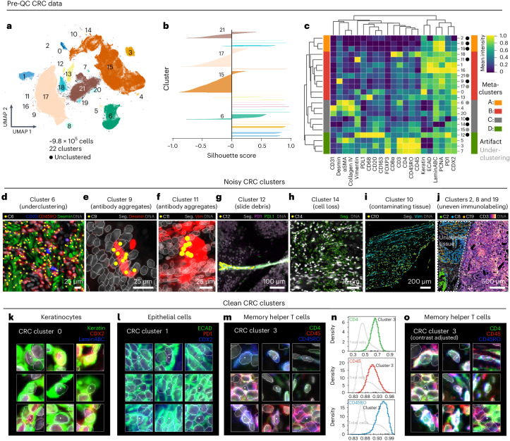
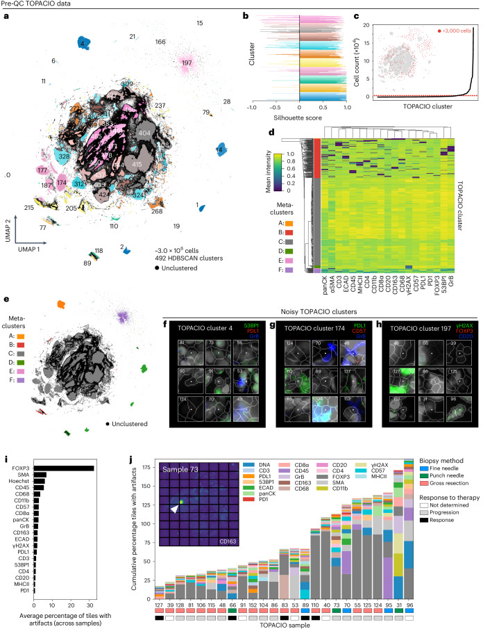
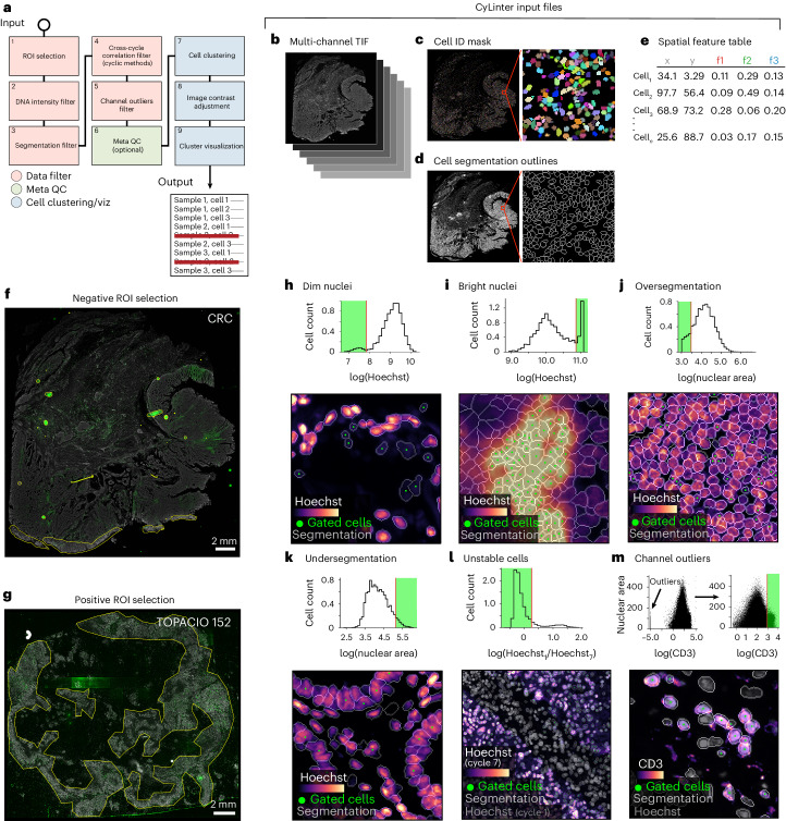
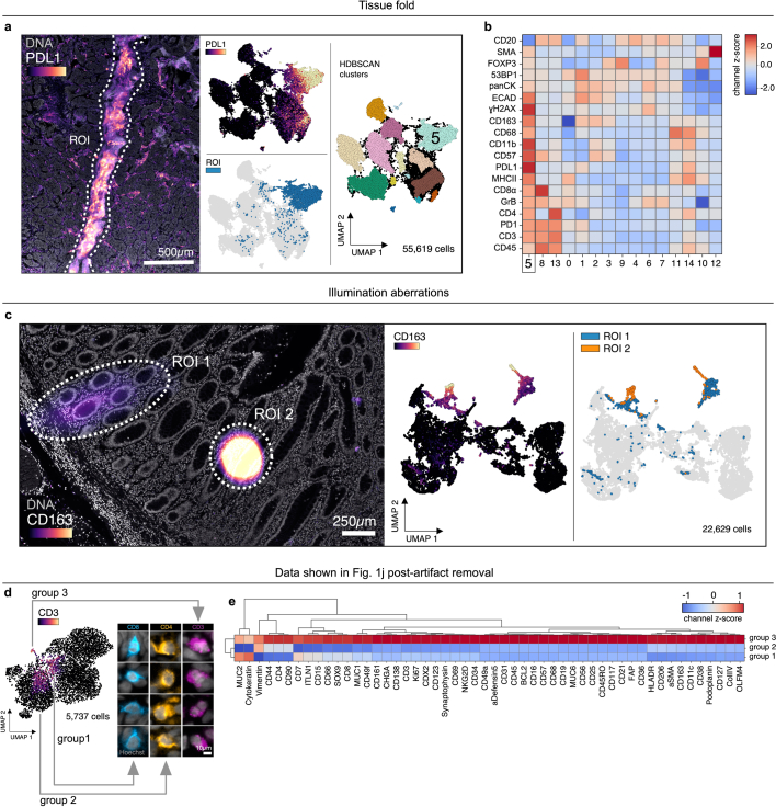
Tumors are complex assemblies of cellular and acellular structures patterned on spatial scales from microns to centimeters. Study of these assemblies has advanced dramatically with the introduction of high-plex spatial profiling. Image-based profiling methods reveal the intensities and spatial distributions of 20-100 proteins at subcellular resolution in 10-10 cells per specimen. Despite extensive work on methods for extracting single-cell data from these images, all tissue images contain artifacts such as folds, debris, antibody aggregates, optical aberrations and image processing errors that arise from imperfections in specimen preparation, data acquisition, image assembly and feature extraction. Here we show that these artifacts dramatically impact single-cell data analysis, obscuring meaningful biological interpretation. We describe an interactive quality control software tool, CyLinter, that identifies and removes data associated with imaging artifacts. CyLinter greatly improves single-cell analysis, especially for archival specimens sectioned many years before data collection, such as those from clinical trials.
肿瘤是细胞和无细胞结构的复杂集合体,其空间尺度从微米到厘米不等。随着高多重空间分析技术的引入,对这些集合体的研究取得了显著进展。基于图像的分析方法能够在亚细胞分辨率下,揭示每个样本中10 - 10个细胞内20 - 100种蛋白质的强度和空间分布。尽管在从这些图像中提取单细胞数据的方法上已经进行了大量工作,但所有组织图像都包含诸如褶皱、碎片、抗体聚集体、光学像差以及在样本制备、数据采集、图像拼接和特征提取过程中由于不完美而产生的图像处理错误等伪像。在这里,我们表明这些伪像会极大地影响单细胞数据分析,模糊有意义的生物学解释。我们描述了一种交互式质量控制软件工具CyLinter,它可以识别并去除与成像伪像相关的数据。CyLinter极大地改进了单细胞分析,特别是对于在数据收集前多年就已切片的存档样本,例如来自临床试验的样本。