• 文献检索
  • 文档翻译
  • 深度研究
  • 学术资讯
  • Suppr Zotero 插件Zotero 插件
  • 邀请有礼
  • 套餐&价格
  • 历史记录
应用&插件
Suppr Zotero 插件Zotero 插件浏览器插件Mac 客户端Windows 客户端微信小程序
定价
高级版会员购买积分包购买API积分包
服务
文献检索文档翻译深度研究API 文档MCP 服务
关于我们
关于 Suppr公司介绍联系我们用户协议隐私条款
关注我们

Suppr 超能文献

核心技术专利:CN118964589B侵权必究
粤ICP备2023148730 号-1Suppr @ 2026

文献检索

告别复杂PubMed语法,用中文像聊天一样搜索,搜遍4000万医学文献。AI智能推荐,让科研检索更轻松。

立即免费搜索

文件翻译

保留排版,准确专业,支持PDF/Word/PPT等文件格式,支持 12+语言互译。

免费翻译文档

深度研究

AI帮你快速写综述,25分钟生成高质量综述,智能提取关键信息,辅助科研写作。

立即免费体验

抗人中间丝蛋白的单克隆抗体。II. 丝蛋白在正常人体组织中的分布。

Monoclonal antibodies to human intermediate filament proteins. II. Distribution of filament proteins in normal human tissues.

作者信息

Gown A M, Vogel A M

出版信息

Am J Pathol. 1984 Feb;114(2):309-21.

PMID:6320650
原文链接:https://pmc.ncbi.nlm.nih.gov/articles/PMC1900342/
Abstract

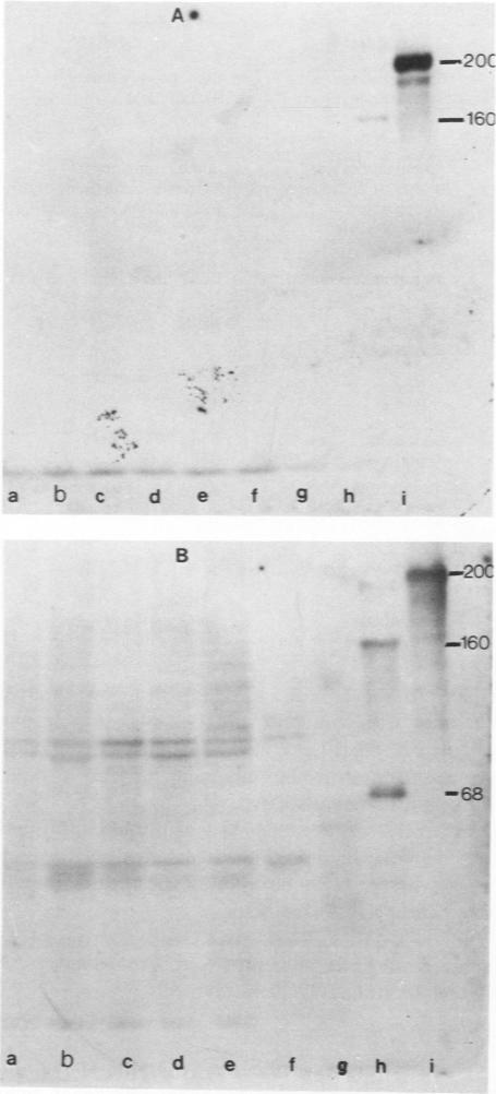
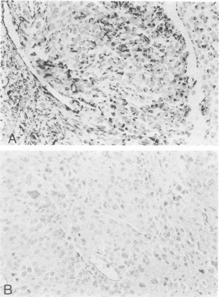
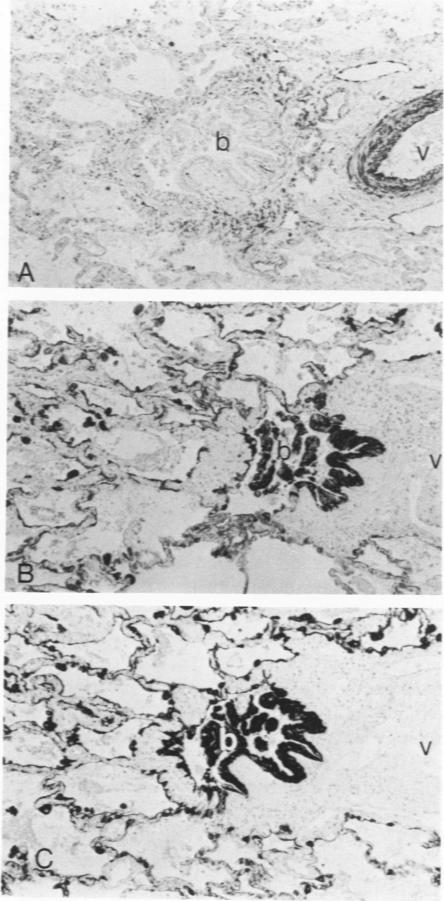
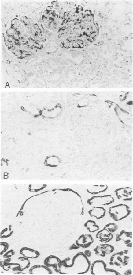
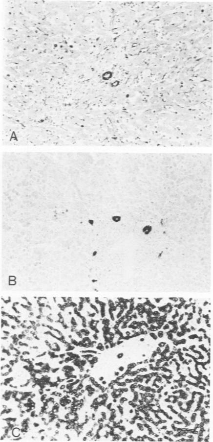
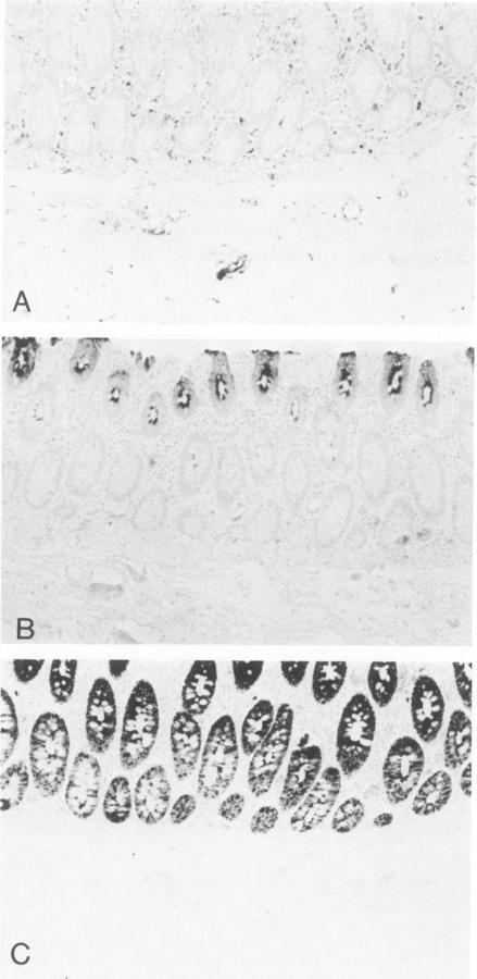
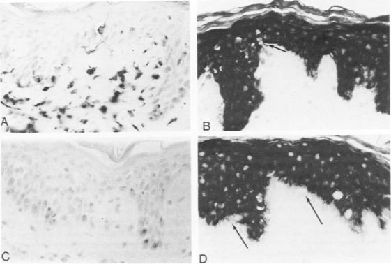
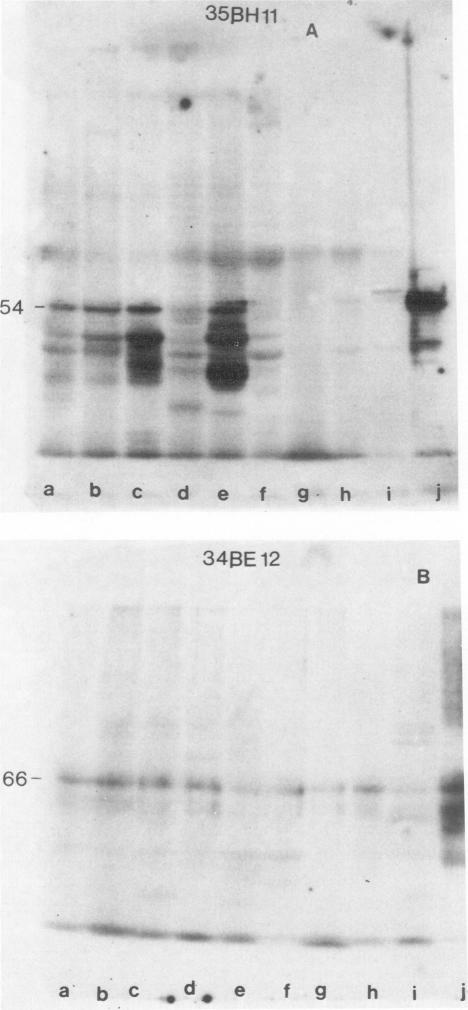
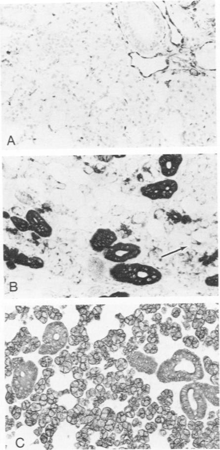
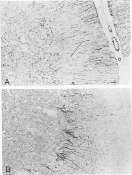
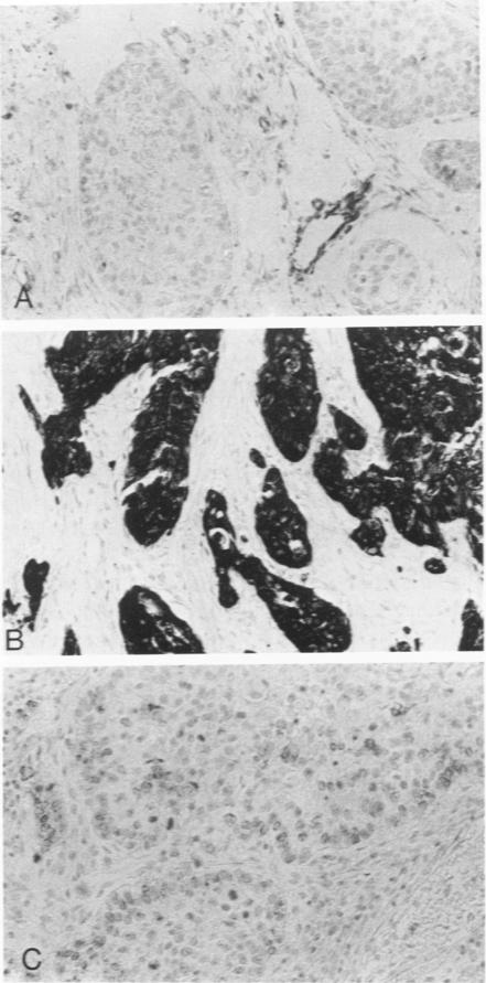
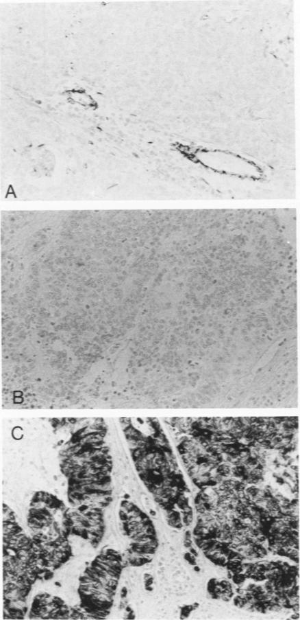
Monoclonal antibodies generated against different human intermediate filament (IF) proteins were assayed on fixed, embedded tissue by the biotin-avidin-immunoperoxidase method for evaluation of the tissue specificity of these antibodies. An antibody (43 beta E8) made to fibroblast IF protein stains mesenchymal tissue such as endothelium, histiocytes, stromal fibroblasts, and Schwann cells but does not stain epithelium, skeletal muscle, lymphocytes, or neurons. Three different anti-cytokeratin antibodies decorate epithelium in three unique patterns. One (35 beta H11) stains all nonsquamous epithelium but fails to recognize squamous epithelium. Antibody 34 beta E12 stains the full thickness of squamous epithelium and ductular epithelium but does not react with hepatocytes, pancreatic acinar cells, proximal renal tubules, or endometrial glands. Antibody 34 beta B4 stains only the suprabasal portion of squamous epithelium. None of these three anti-cytokeratin antibodies reacts with nerve or mesenchymal tissue. Two anti-neurofilament antibodies recognize only neurons, failing to react with epithelial or mesenchymal tissue. We conclude that these anti-intermediate filament antibodies can be used as tissue-specific markers. Neoplasms retain the same intermediate filament patterns as the normal parental tissue; therefore, these antibodies can be used as diagnostic aids in surgical pathology.

摘要

采用生物素-抗生物素蛋白-免疫过氧化物酶法,在固定、包埋的组织上检测针对不同人类中间丝(IF)蛋白产生的单克隆抗体,以评估这些抗体的组织特异性。一种针对成纤维细胞IF蛋白产生的抗体(43βE8)可使间充质组织如内皮细胞、组织细胞、基质成纤维细胞和施万细胞染色,但不能使上皮细胞、骨骼肌、淋巴细胞或神经元染色。三种不同的抗细胞角蛋白抗体以三种独特的模式使上皮细胞染色。一种(35βH11)可使所有非鳞状上皮细胞染色,但不能识别鳞状上皮细胞。抗体34βE12可使鳞状上皮细胞和导管上皮细胞全层染色,但不与肝细胞、胰腺腺泡细胞、近端肾小管或子宫内膜腺反应。抗体34βB4仅使鳞状上皮细胞的基底上层部分染色。这三种抗细胞角蛋白抗体均不与神经组织或间充质组织反应。两种抗神经丝抗体仅识别神经元,不与上皮组织或间充质组织反应。我们得出结论,这些抗中间丝抗体可作为组织特异性标志物。肿瘤保留与正常亲代组织相同的中间丝模式;因此,这些抗体可作为外科病理学中的诊断辅助工具。

https://cdn.ncbi.nlm.nih.gov/pmc/blobs/3fd7/1900342/26c8f315c618/amjpathol00185-0138-a.jpg
https://cdn.ncbi.nlm.nih.gov/pmc/blobs/3fd7/1900342/4aed608af0db/amjpathol00185-0129-a.jpg
https://cdn.ncbi.nlm.nih.gov/pmc/blobs/3fd7/1900342/7e4f150175f6/amjpathol00185-0129-b.jpg
https://cdn.ncbi.nlm.nih.gov/pmc/blobs/3fd7/1900342/1e1bc4cb0d8e/amjpathol00185-0131-a.jpg
https://cdn.ncbi.nlm.nih.gov/pmc/blobs/3fd7/1900342/536eacb3a270/amjpathol00185-0131-b.jpg
https://cdn.ncbi.nlm.nih.gov/pmc/blobs/3fd7/1900342/000f42b410c0/amjpathol00185-0132-a.jpg
https://cdn.ncbi.nlm.nih.gov/pmc/blobs/3fd7/1900342/acdf56fd6fb2/amjpathol00185-0133-a.jpg
https://cdn.ncbi.nlm.nih.gov/pmc/blobs/3fd7/1900342/773ae4e5046e/amjpathol00185-0134-a.jpg
https://cdn.ncbi.nlm.nih.gov/pmc/blobs/3fd7/1900342/70261dccd42a/amjpathol00185-0134-b.jpg
https://cdn.ncbi.nlm.nih.gov/pmc/blobs/3fd7/1900342/d7bdafa2469d/amjpathol00185-0135-a.jpg
https://cdn.ncbi.nlm.nih.gov/pmc/blobs/3fd7/1900342/fc2b688eacfa/amjpathol00185-0135-b.jpg
https://cdn.ncbi.nlm.nih.gov/pmc/blobs/3fd7/1900342/072308198d7e/amjpathol00185-0137-a.jpg
https://cdn.ncbi.nlm.nih.gov/pmc/blobs/3fd7/1900342/aafac8ccd769/amjpathol00185-0137-b.jpg
https://cdn.ncbi.nlm.nih.gov/pmc/blobs/3fd7/1900342/26c8f315c618/amjpathol00185-0138-a.jpg

相似文献

1
Monoclonal antibodies to human intermediate filament proteins. II. Distribution of filament proteins in normal human tissues.抗人中间丝蛋白的单克隆抗体。II. 丝蛋白在正常人体组织中的分布。
Am J Pathol. 1984 Feb;114(2):309-21.
2
Monoclonal antibodies to intermediate filament proteins of human cells: unique and cross-reacting antibodies.针对人类细胞中间丝蛋白的单克隆抗体:独特抗体与交叉反应抗体
J Cell Biol. 1982 Nov;95(2 Pt 1):414-24. doi: 10.1083/jcb.95.2.414.
3
Monoclonal antibodies to human intermediate filament proteins. III. Analysis of tumors.
Am J Clin Pathol. 1985 Oct;84(4):413-24. doi: 10.1093/ajcp/84.4.413.
4
Immunohistochemical detection of intermediate filament proteins in formalin fixed normal and neoplastic canine tissues.福尔马林固定的正常和肿瘤性犬组织中中间丝蛋白的免疫组织化学检测
Can J Vet Res. 1990 Jun;54(3):360-5.
5
Antibodies to intermediate filament proteins as molecular markers in clinical tumor pathology. Differentiation of carcinomas by their reaction with different cytokeratin antibodies.中间丝蛋白抗体作为临床肿瘤病理学中的分子标志物。通过癌与不同细胞角蛋白抗体的反应对癌进行鉴别。
Pathol Res Pract. 1982;175(2-3):212-26. doi: 10.1016/S0344-0338(82)80109-X.
6
Application of monoclonal antibodies to intermediate filament proteins in surgical pathology of head and neck tumours. An overview.
Appl Pathol. 1988;6(1):35-48.
7
Expression of intermediate filament proteins in fetal and adult human kidney: modulations of intermediate filament patterns during development and in damaged tissue.中间丝蛋白在胎儿及成人肾脏中的表达:发育过程中和受损组织中中间丝模式的调节
Lab Invest. 1991 Jul;65(1):74-86.
8
Coexpression of intermediate filament polypeptides in human fetal and adult tissues.中间丝多肽在人类胎儿和成人组织中的共表达。
Lab Invest. 1987 Oct;57(4):359-69.
9
[Immunohistochemical study of human breast tumors using monoclonal antibodies to intermediate filament proteins (nonproliferating epithelial structures in breast dysplasia)].
Arkh Patol. 1985;47(9):53-9.
10
Detection of epithelial- and neural type of intermediate filament proteins in human lung tumors.人肺肿瘤中上皮型和神经型中间丝蛋白的检测
Acta Histochem Suppl. 1987;34:45-56.

引用本文的文献

1
Xanthogranulomatous epithelial tumor: report of 6 cases of a novel, potentially deceptive lesion with a predilection for young women.黄肉芽肿性上皮瘤:6 例新型、具有潜在欺骗性病变的报告,好发于年轻女性。
Mod Pathol. 2020 Oct;33(10):1889-1895. doi: 10.1038/s41379-020-0562-8. Epub 2020 May 15.
2
Diagnostic Biomarkers in Oral Verrucous Carcinoma: A Systematic Review.口腔疣状癌的诊断生物标志物:一项系统综述。
Pathol Oncol Res. 2017 Jan;23(1):19-32. doi: 10.1007/s12253-016-0150-x. Epub 2016 Dec 6.
3
Tumor necrosis factor-α confers cardioprotection through ectopic expression of keratins K8 and K18.

本文引用的文献

1
Immunohistochemical localization of keratin in normal human tissues.角蛋白在正常人体组织中的免疫组织化学定位。
Lab Invest. 1980 Jan;42(1):91-6.
2
The catalog of human cytokeratins: patterns of expression in normal epithelia, tumors and cultured cells.人类细胞角蛋白目录:在正常上皮、肿瘤及培养细胞中的表达模式
Cell. 1982 Nov;31(1):11-24. doi: 10.1016/0092-8674(82)90400-7.
3
Immunolocalization of keratin polypeptides in human epidermis using monoclonal antibodies.使用单克隆抗体对人表皮中的角蛋白多肽进行免疫定位。
肿瘤坏死因子-α通过角蛋白K8和K18的异位表达赋予心脏保护作用。
Nat Med. 2015 Sep;21(9):1076-84. doi: 10.1038/nm.3925. Epub 2015 Aug 17.
4
PI3K/AKT/mTOR pathway plays a major pathogenetic role in glycogen accumulation and tumor development in renal distal tubules of rats and men.PI3K/AKT/mTOR信号通路在大鼠和人类肾远端小管的糖原积累及肿瘤发生发展中起主要致病作用。
Oncotarget. 2015 May 30;6(15):13036-48. doi: 10.18632/oncotarget.3675.
5
p63 is more sensitive and specific than 34βE12 to differentiate adenocarcinoma of prostate from cancer mimickers.p63 比 34βE12 更敏感和特异,可用于鉴别前列腺腺癌与癌类似物。
Iran J Basic Med Sci. 2014 Jul;17(7):497-501.
6
Development of epithelial and mesenchymal regionalization of the human fetal utero-vaginal anlagen.人胎儿子宫阴道原基的上皮和中胚层区域化的发展。
J Anat. 2013 Apr;222(4):462-72. doi: 10.1111/joa.12029. Epub 2013 Feb 13.
7
Plakophilin-2: a cell-cell adhesion plaque molecule of selective and fundamental importance in cardiac functions and tumor cell growth. plakophilin-2:一种细胞间黏附斑分子,在心功能和肿瘤细胞生长中具有选择性和重要的基础作用。
Cell Tissue Res. 2012 May;348(2):281-94. doi: 10.1007/s00441-011-1314-3. Epub 2012 Jan 28.
8
Immunocytochemical analysis for differential diagnosis of thyroid lesions using liquid-based cytology.使用液基细胞学进行甲状腺病变鉴别诊断的免疫细胞化学分析。
Nagoya J Med Sci. 2011 Feb;73(1-2):15-24.
9
Evaluation of routine application of P504S, 34betaE12 and p63 immunostaining on 250 prostate needle biopsy specimens.评估 P504S、34βE12 和 p63 免疫染色在 250 例前列腺针芯活检标本中的常规应用。
Int Urol Nephrol. 2010 Jun;42(2):325-30. doi: 10.1007/s11255-009-9622-1. Epub 2009 Aug 5.
10
Ebp1 expression in benign and malignant prostate.Ebp1在良性和恶性前列腺中的表达。
Cancer Cell Int. 2008 Nov 24;8:18. doi: 10.1186/1475-2867-8-18.
J Cell Biol. 1982 Nov;95(2 Pt 1):580-8. doi: 10.1083/jcb.95.2.580.
4
Monoclonal antibodies to intermediate filament proteins of human cells: unique and cross-reacting antibodies.针对人类细胞中间丝蛋白的单克隆抗体:独特抗体与交叉反应抗体
J Cell Biol. 1982 Nov;95(2 Pt 1):414-24. doi: 10.1083/jcb.95.2.414.
5
Correlation of specific keratins with different types of epithelial differentiation: monoclonal antibody studies.特定角蛋白与不同类型上皮分化的相关性:单克隆抗体研究
Cell. 1982 Sep;30(2):361-72. doi: 10.1016/0092-8674(82)90234-3.
6
Color modification of diaminobenzidine (DAB) precipitation by metallic ions and its application for double immunohistochemistry.金属离子对二氨基联苯胺(DAB)沉淀的颜色修饰及其在双重免疫组织化学中的应用。
J Histochem Cytochem. 1982 Oct;30(10):1079-82. doi: 10.1177/30.10.6182185.
7
Monoclonal antibodies provide specific intramolecular markers for the study of epithelial tonofilament organization.单克隆抗体为上皮张力丝组织的研究提供了特异性的分子内标记。
J Cell Biol. 1982 Mar;92(3):665-73. doi: 10.1083/jcb.92.3.665.
8
Antibodies to different intermediate filament proteins. Cell type-specific markers on paraffin-embedded human tissues.针对不同中间丝蛋白的抗体。石蜡包埋的人体组织上的细胞类型特异性标志物。
Lab Invest. 1981 Nov;45(5):427-34.
9
Immunochemical identification of intermediate-sized filaments in human neoplastic cells. A diagnostic aid for the surgical pathologist.人肿瘤细胞中中间丝的免疫化学鉴定。对外科病理学家的诊断辅助。
Am J Pathol. 1981 Sep;104(3):206-16.
10
Use of avidin-biotin-peroxidase complex (ABC) in immunoperoxidase techniques: a comparison between ABC and unlabeled antibody (PAP) procedures.抗生物素蛋白-生物素-过氧化物酶复合物(ABC)在免疫过氧化物酶技术中的应用:ABC法与未标记抗体(PAP)法的比较。
J Histochem Cytochem. 1981 Apr;29(4):577-80. doi: 10.1177/29.4.6166661.