• 文献检索
  • 文档翻译
  • 深度研究
  • 学术资讯
  • Suppr Zotero 插件Zotero 插件
  • 邀请有礼
  • 套餐&价格
  • 历史记录
应用&插件
Suppr Zotero 插件Zotero 插件浏览器插件Mac 客户端Windows 客户端微信小程序
定价
高级版会员购买积分包购买API积分包
服务
文献检索文档翻译深度研究API 文档MCP 服务
关于我们
关于 Suppr公司介绍联系我们用户协议隐私条款
关注我们

Suppr 超能文献

核心技术专利:CN118964589B侵权必究
粤ICP备2023148730 号-1Suppr @ 2026

文献检索

告别复杂PubMed语法,用中文像聊天一样搜索,搜遍4000万医学文献。AI智能推荐,让科研检索更轻松。

立即免费搜索

文件翻译

保留排版,准确专业,支持PDF/Word/PPT等文件格式,支持 12+语言互译。

免费翻译文档

深度研究

AI帮你快速写综述,25分钟生成高质量综述,智能提取关键信息,辅助科研写作。

立即免费体验

海德堡视网膜断层扫描仪II测定的视盘形态与开角型青光眼患者视功能之间的相关性

Correlation between morphology of optic disc determined by Heidelberg Retina Tomograph II and visual function in eyes with open-angle glaucoma.

作者信息

Omodaka Kazuko, Nakazawa Toru, Otomo Takaaki, Nakamura Masahiko, Fuse Nobuo, Nishida Kohji

机构信息

Department of Ophthalmology, Tohoku University Graduate School of Medicine, 1-1 Seiryo-machi, Aoba-ku, Sendai, Miyagi 980-8574, Japan.

出版信息

Clin Ophthalmol. 2010 Jul 30;4:765-72. doi: 10.2147/opth.s9741.

DOI:10.2147/opth.s9741
PMID:20689792
原文链接:https://pmc.ncbi.nlm.nih.gov/articles/PMC2915862/
Abstract

PURPOSE

To investigate the correlation between the morphology of the optic disc and the visual function of eyes with open-angle glaucoma (OAG).

PATIENTS AND METHODS

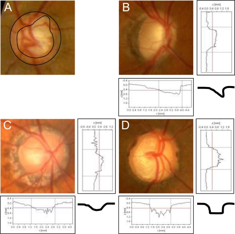
One hundred and three eyes of 76 patients with OAG were studied. The baseline optic disc morphology was used to classify the eyes into four types: focal ischemic type (FI), myopic glaucomatous type (MY), senile sclerotic type (SS), and generalized enlargement type (GE). The morphological parameters of the disc were determined by the Heidelberg Retina Tomograph II (HRT-II) and the visual function by the mean deviation (MD) of the Humphrey field analyzer.

RESULTS

Fourteen eyes were classified as the FI type; 52 as the MY type; 17 as the SS type; and 20 as the GE type. The highest correlation coefficients of HRT-II parameters to the MDs was the cup/disc area ratio (r = -0.27) for all groups, the vertical cup/disc ratio (r = -0.42) in the MY group, the maximum cup depth (r = 0.49) in the SS group, and the cup area (r = -0.70) in the GE group. However, none of the parameters was correlated in the FI group.

CONCLUSIONS

The correlation between the HRT-II parameters and MDs was different for the four disc types. These findings suggest that a classification of optic disc morphology had a benefit for interpreting the measured values of HRT-II.

摘要

目的

研究开角型青光眼(OAG)患者视盘形态与视功能之间的相关性。

患者与方法

对76例OAG患者的103只眼睛进行了研究。根据基线视盘形态将眼睛分为四种类型:局灶缺血型(FI)、近视性青光眼型(MY)、老年性硬化型(SS)和普遍性扩大型(GE)。视盘的形态学参数由海德堡视网膜断层扫描仪II(HRT-II)测定,视功能由 Humphrey 视野分析仪的平均偏差(MD)测定。

结果

14只眼睛被分类为FI型;52只为MY型;17只为SS型;20只为GE型。所有组中,HRT-II参数与MD的最高相关系数为杯盘面积比(r = -0.27),MY组为垂直杯盘比(r = -0.42),SS组为最大杯深(r = 0.49),GE组为杯面积(r = -0.70)。然而,FI组中没有参数具有相关性。

结论

四种视盘类型中,HRT-II参数与MD之间的相关性有所不同。这些发现表明,视盘形态分类有助于解释HRT-II的测量值。

https://cdn.ncbi.nlm.nih.gov/pmc/blobs/4a5b/2915862/1bfeeb21526a/opth-4-765f1.jpg

相似文献

1
Correlation between morphology of optic disc determined by Heidelberg Retina Tomograph II and visual function in eyes with open-angle glaucoma.海德堡视网膜断层扫描仪II测定的视盘形态与开角型青光眼患者视功能之间的相关性
Clin Ophthalmol. 2010 Jul 30;4:765-72. doi: 10.2147/opth.s9741.
2
Characteristic correlations of the structure-function relationship in different glaucomatous disc types.不同青光眼视盘类型中结构-功能关系的特征性关联
Jpn J Ophthalmol. 2015 Jul;59(4):223-9. doi: 10.1007/s10384-015-0379-z. Epub 2015 Apr 11.
3
OCT-Based Quantification and Classification of Optic Disc Structure in Glaucoma Patients.基于光学相干断层扫描的青光眼患者视盘结构定量与分类
PLoS One. 2016 Aug 24;11(8):e0160226. doi: 10.1371/journal.pone.0160226. eCollection 2016.
4
Optic disc topography and peripapillary retinal nerve fiber layer thickness in nonarteritic ischemic optic neuropathy and open-angle glaucoma.非动脉炎性缺血性视神经病变和开角型青光眼中的视盘地形图及视乳头周围视网膜神经纤维层厚度
Ophthalmology. 2006 Aug;113(8):1340-4. doi: 10.1016/j.ophtha.2006.01.072. Epub 2006 Jun 22.
5
Correlation of disc morphology quantified on stereophotographs to results by Heidelberg Retina Tomograph II, GDx variable corneal compensation, and visual field tests.立体照片定量分析盘形态与海德堡视网膜断层扫描仪 II、GDx 可变角膜补偿和视野检查结果的相关性。
Ophthalmology. 2010 Feb;117(2):282-9. doi: 10.1016/j.ophtha.2009.07.004. Epub 2009 Dec 6.
6
Morphological features and important parameters of large optic discs for diagnosing glaucoma.用于诊断青光眼的大视盘的形态学特征及重要参数。
PLoS One. 2015 Mar 23;10(3):e0118920. doi: 10.1371/journal.pone.0118920. eCollection 2015.
7
Correlation between corneal thickness and optic disc morphology in normal tension glaucoma using modern technical analysis.运用现代技术分析研究正常眼压性青光眼中角膜厚度与视盘形态之间的相关性。
Rom J Morphol Embryol. 2014;55(3):857-62.
8
Glaucoma diagnostic accuracy of optical coherence tomography parameters in early glaucoma with different types of optic disc damage.光学相干断层扫描参数对不同类型视盘损伤早期青光眼的诊断准确性。
Ophthalmology. 2014 Oct;121(10):1990-7. doi: 10.1016/j.ophtha.2014.04.030. Epub 2014 Jun 14.
9
Evaluation of optic nerve head using a newly developed stereo retinal imaging technique by glaucoma specialist and non-expert-certified orthoptist.使用由青光眼专家和未经认证的视轴矫正专家新开发的立体视网膜成像技术评估视神经头。
J Glaucoma. 2013 Dec;22(9):698-706. doi: 10.1097/IJG.0b013e318264be18.
10
Progression of visual field defects in eyes with different optic disc appearances in patients with normal tension glaucoma.正常眼压性青光眼患者不同视盘外观眼中视野缺损的进展。
J Glaucoma. 2012 Aug;21(6):426-30. doi: 10.1097/IJG.0b013e3182182897.

引用本文的文献

1
OCT-Based Quantification and Classification of Optic Disc Structure in Glaucoma Patients.基于光学相干断层扫描的青光眼患者视盘结构定量与分类
PLoS One. 2016 Aug 24;11(8):e0160226. doi: 10.1371/journal.pone.0160226. eCollection 2016.
2
Clinical Factors Associated with Lamina Cribrosa Thickness in Patients with Glaucoma, as Measured with Swept Source Optical Coherence Tomography.扫频源光学相干断层扫描测量青光眼患者筛板厚度的相关临床因素
PLoS One. 2016 Apr 21;11(4):e0153707. doi: 10.1371/journal.pone.0153707. eCollection 2016.
3
3D evaluation of the lamina cribrosa with swept-source optical coherence tomography in normal tension glaucoma.

本文引用的文献

1
Ability of the heidelberg retina tomograph to detect early glaucomatous visual field loss.海德堡视网膜断层扫描仪检测早期青光眼视野损失的能力。
J Glaucoma. 1995 Aug;4(4):242-7.
2
Laser scanning tomography of optic discs of the normal Japanese population in a population-based setting.基于人群的日本正常人群视盘的激光扫描断层扫描
Ophthalmology. 2009 Feb;116(2):223-30. doi: 10.1016/j.ophtha.2008.09.013. Epub 2008 Dec 12.
3
Monitoring glaucoma progression.监测青光眼进展。
正常眼压性青光眼中使用扫频源光学相干断层扫描对筛板进行三维评估。
PLoS One. 2015 Apr 15;10(4):e0122347. doi: 10.1371/journal.pone.0122347. eCollection 2015.
4
Characteristic correlations of the structure-function relationship in different glaucomatous disc types.不同青光眼视盘类型中结构-功能关系的特征性关联
Jpn J Ophthalmol. 2015 Jul;59(4):223-9. doi: 10.1007/s10384-015-0379-z. Epub 2015 Apr 11.
5
Stereoscopic analysis of optic nerve head parameters in primary open angle glaucoma: the glaucoma stereo analysis study.原发性开角型青光眼视神经乳头参数的立体分析:青光眼立体分析研究
PLoS One. 2014 Jun 12;9(6):e99138. doi: 10.1371/journal.pone.0099138. eCollection 2014.
6
Association between optic nerve blood flow and objective examinations in glaucoma patients with generalized enlargement disc type.视盘普遍增大型青光眼患者视神经血流与客观检查之间的关联
Clin Ophthalmol. 2011;5:1549-56. doi: 10.2147/OPTH.S22097. Epub 2011 Oct 28.
Prog Brain Res. 2008;173:59-73. doi: 10.1016/S0079-6123(08)01106-0.
4
Heidelberg retina tomograph parameters of the optic disc in eyes with progressive retinal nerve fibre layer defects.患有进行性视网膜神经纤维层缺损的眼睛视盘的海德堡视网膜断层扫描参数。
Acta Ophthalmol. 2008 Sep;86(6):603-8. doi: 10.1111/j.1600-0420.2007.01119.x.
5
Prevalence and risk factors for refractive errors in the Singapore Malay Eye Survey.新加坡马来人眼研究中屈光不正的患病率及危险因素
Ophthalmology. 2008 Oct;115(10):1713-9. doi: 10.1016/j.ophtha.2008.03.016. Epub 2008 May 16.
6
Analysis of HRT images: comparison of reference planes.HRT图像分析:参考平面的比较
Invest Ophthalmol Vis Sci. 2008 Sep;49(9):3970-5. doi: 10.1167/iovs.08-1764. Epub 2008 May 9.
7
Relationship between Humphrey 30-2 SITA Standard Test, Matrix 30-2 threshold test, and Heidelberg retina tomograph in ocular hypertensive and glaucoma patients.高眼压症和青光眼患者中 Humphrey 30-2 SITA 标准测试、Matrix 30-2 阈值测试与海德堡视网膜断层扫描之间的关系。
J Glaucoma. 2008 Apr-May;17(3):203-10. doi: 10.1097/IJG.0b013e31815a3493.
8
Refractive errors in an elderly Japanese population: the Tajimi study.日本老年人群中的屈光不正:多治见研究
Ophthalmology. 2008 Feb;115(2):363-370.e3. doi: 10.1016/j.ophtha.2007.03.075.
9
Optic disk and nerve fiber layer imaging to detect glaucoma.用于检测青光眼的视盘和神经纤维层成像
Am J Ophthalmol. 2007 Nov;144(5):724-32. doi: 10.1016/j.ajo.2007.07.010. Epub 2007 Sep 14.
10
Evaluation of optical coherence tomography and heidelberg retinal tomography parameters in detecting early and moderate glaucoma.光学相干断层扫描和海德堡视网膜断层扫描参数在检测早期和中度青光眼方面的评估
Invest Ophthalmol Vis Sci. 2007 Jul;48(7):3138-45. doi: 10.1167/iovs.06-1407.