Department of Biomedical Engineering, University of Michigan, Ann Arbor, Michigan 48109-2099, USA.
J Control Release. 2012 Jan 10;157(1):103-11. doi: 10.1016/j.jconrel.2011.09.068. Epub 2011 Sep 16.
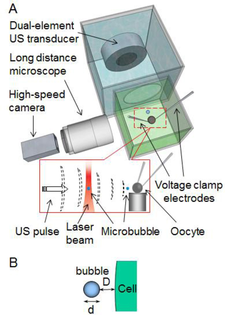
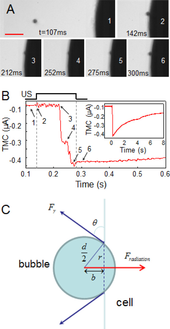
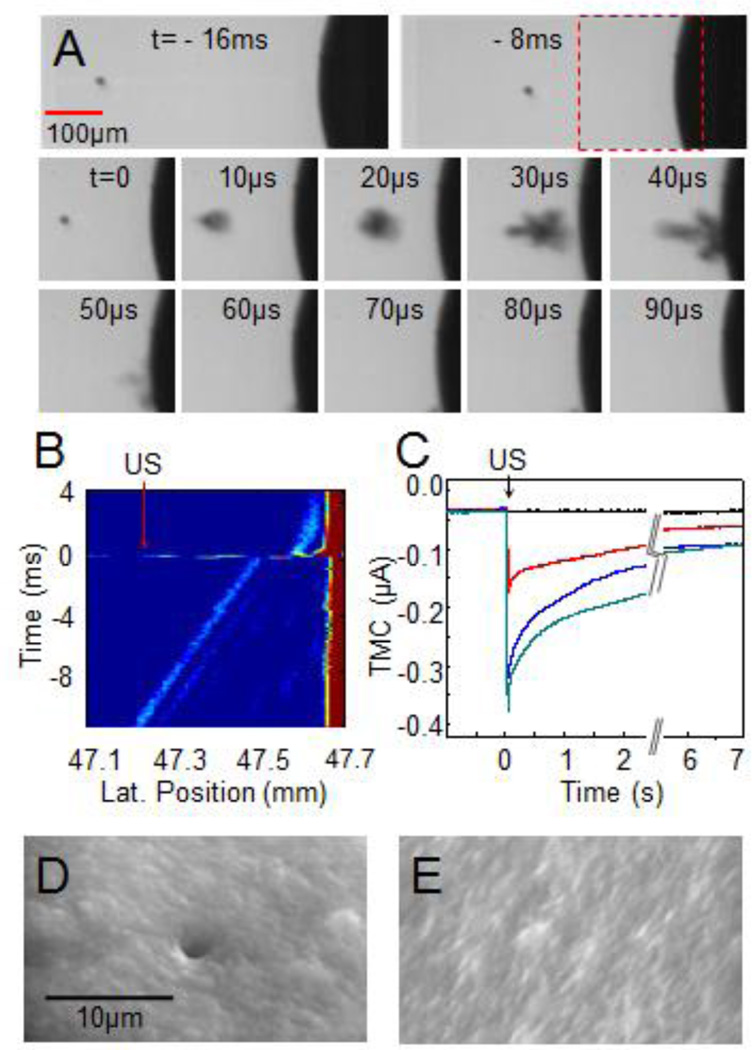
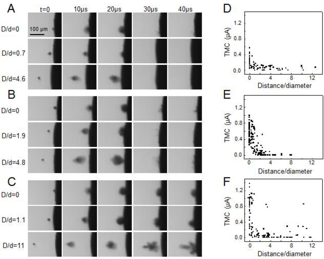
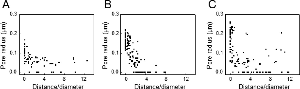
Sonoporation is the membrane disruption generated by ultrasound and has been exploited as a non-viral strategy for drug and gene delivery. Acoustic cavitation of microbubbles has been recognized to play an important role in sonoporation. However, due to the lack of adequate techniques for precise control of cavitation activities and real-time assessment of the resulting sub-micron process of sonoporation, limited knowledge has been available regarding the detail processes and correlation of cavitation with membrane disruption at the single cell level. In the current study, we developed a combined approach including optical, acoustical, and electrophysiological techniques to enable synchronized manipulation, imaging, and measurement of cavitation of single bubbles and the resulting cell membrane disruption in real-time. Using a self-focused femtosecond laser and high frequency ultrasound (7.44MHz) pulses, a single microbubble was generated and positioned at a desired distance from the membrane of a Xenopus oocyte. Cavitation of the bubble was achieved by applying a low frequency (1.5MHz) ultrasound pulse (duration 13.3 or 40μs) to induce bubble collapse. Disruption of the cell membrane was assessed by the increase in the transmembrane current (TMC) of the cell under voltage clamp. Simultaneous high-speed bright field imaging of cavitation and measurements of the TMC were obtained to correlate the ultrasound-generated bubble activities with the cell membrane poration. The change in membrane permeability was directly associated with the formation of a sub-micrometer pore from a local membrane rupture generated by bubble collapse or bubble compression depending on ultrasound amplitude and duration. The impact of the bubble collapse on membrane permeation decreased rapidly with increasing distance (D) between the bubble (diameter d) and the cell membrane. The effective range of cavitation impact on membrane poration was determined to be D/d=0.75. The maximum mean radius of the pores was estimated from the measured TMC to be 0.106±0.032μm (n=70) for acoustic pressure of 1.5MPa (duration 13.3μs), and increased to 0.171±0.030μm (n=125) for acoustic pressure of 1.7MPa and to 0.182±0.052μm (n=112) for a pulse duration of 40μs (1.5MPa). These results from controlled cell membrane permeation by cavitation of single bubbles revealed insights and key factors affecting sonoporation at the single cell level.
声致孔作用是超声产生的膜破裂作用,已被用作药物和基因传递的非病毒策略。微泡的声空化作用已被认为在声致孔作用中起着重要作用。然而,由于缺乏精确控制空化活动和实时评估亚微米级声致孔过程的充分技术,因此对于在单细胞水平上的空化与膜破裂之间的细节过程和相关性的了解有限。在本研究中,我们开发了一种结合光学、声学和电生理学技术的方法,以实现对单个气泡的空化作用及其对细胞膜的实时破坏的同步操纵、成像和测量。使用自聚焦飞秒激光和高频超声(7.44MHz)脉冲,生成单个微泡并将其放置在距非洲爪蟾卵母细胞膜的期望距离处。通过施加低频(1.5MHz)超声脉冲(持续时间 13.3 或 40μs)来实现气泡的空化作用,以诱导气泡的崩溃。通过细胞膜电压钳下细胞跨膜电流(TMC)的增加来评估细胞膜的破坏。同时获得了空化作用的高速亮场成像和 TMC 的测量结果,以将超声产生的气泡活动与细胞膜穿孔相关联。膜通透性的变化直接与局部膜破裂产生的亚微米孔的形成有关,局部膜破裂是由气泡的崩溃或气泡的压缩引起的,具体取决于超声幅度和持续时间。气泡崩溃对膜渗透的影响随着气泡(直径 d)与细胞膜之间距离(D)的增加而迅速减小。通过测量跨膜电流(TMC)来估计孔的有效半径,对于 1.5MPa(持续时间 13.3μs)的声压,平均最大半径为 0.106±0.032μm(n=70),对于 1.7MPa 的声压增加到 0.171±0.030μm(n=125),对于持续时间为 40μs(1.5MPa)增加到 0.182±0.052μm(n=112)。这些通过单个气泡的空化作用控制细胞膜通透性的结果揭示了影响单细胞水平声致孔作用的见解和关键因素。