Kernt Marcus, Cheuteu Raoul E, Cserhati Sarah, Seidensticker Florian, Liegl Raffael G, Lang Julian, Haritoglou Christos, Kampik Anselm, Ulbig Michael W, Neubauer Aljoscha S
Department of Ophthalmology, Ludwig Maximilian University of Munich, Germany.
Clin Ophthalmol. 2012;6:289-96. doi: 10.2147/OPTH.S27859. Epub 2012 Feb 27.
To investigate treatment-related pain and the accuracy of navigated laser photocoagulation in the treatment of clinically significant macular edema.
Focal laser treatment of diabetic macular edema in 54 consecutive patients was digitally planned on fundus images and performed using the navigated laser photocoagulation system Navilas(®) (OD-OS GmbH, Teltow, Germany). Treatment-related pain was quantified on a visual analog scale directly after treatment and compared with a matched control group who received conventional laser treatment (n = 46). In addition, for Navilas-treated patients, the accuracy of spot placement on color images was analyzed 1 month after treatment.
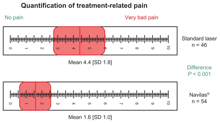
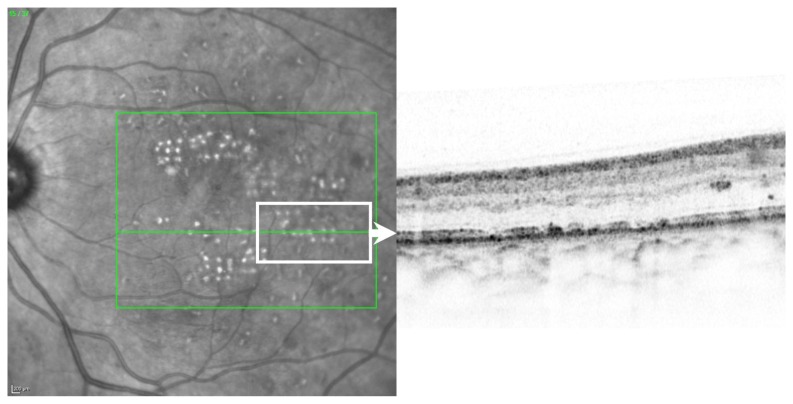
In total, 5423 laser spots (mean 100 per eye) were analyzed. With navigated treatment, 90% of laser spots were visible on color images, of which 96% were within 100 μm from the target. Eighty percent of the laser spots were placed and visible within the 100 μm target on an intention-to-treat basis for color imaging. Optical coherence topography confirmed that laser effects were limited to the outer retina. Treatment-related pain following navigated laser photocoagulation was significantly lower than that of conventional laser treatment (1.6 vs 4.4 on a visual analog scale, P < 0.001).
Navigated laser effects could be visualized to a high percentage on post-treatment color images, and their location showed a high concordance to targeted areas. Patients reported that treatment-related pain following Navilas laser photocoagulation was significantly lower than pain following conventional laser treatment.
研究与治疗相关的疼痛以及导航激光光凝术治疗临床显著性黄斑水肿的准确性。
对54例连续性糖尿病性黄斑水肿患者的局灶性激光治疗在眼底图像上进行数字规划,并使用导航激光光凝系统Navilas®(德国特尔托夫OD-OS有限公司)实施。治疗后即刻采用视觉模拟评分法对与治疗相关的疼痛进行量化,并与接受传统激光治疗的匹配对照组(n = 46)进行比较。此外,对于接受Navilas治疗的患者,在治疗后1个月分析彩色图像上光斑放置的准确性。
共分析了5423个激光光斑(平均每只眼100个)。采用导航治疗时,90%的激光光斑在彩色图像上可见,其中96%距离目标在100μm以内。在彩色成像的意向性分析中,80%的激光光斑放置在100μm目标范围内且可见。光学相干断层扫描证实激光效应局限于视网膜外层。导航激光光凝术后与治疗相关的疼痛显著低于传统激光治疗(视觉模拟评分为1.6 vs 4.4,P < 0.001)。
治疗后彩色图像上可高比例地显示导航激光效应,其位置与目标区域高度一致。患者报告称,Navilas激光光凝术后与治疗相关的疼痛显著低于传统激光治疗后的疼痛。