Institute of Physics, Academia Sinica, Nankang, Taipei 115, Taiwan.
J Nanobiotechnology. 2012 Mar 12;10:10. doi: 10.1186/1477-3155-10-10.
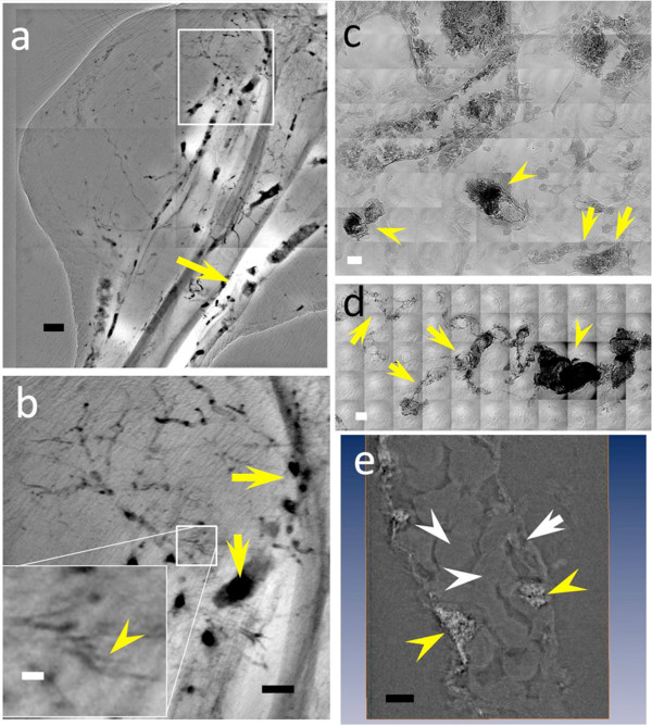
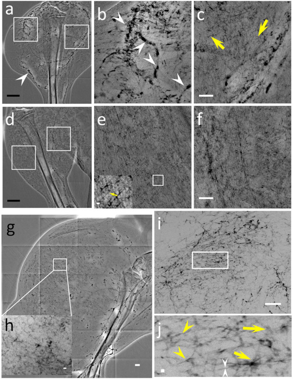
Angiogenesis is widely investigated in conjunction with cancer development, in particular because of the possibility of early stage detection and of new therapeutic strategies. However, such studies are negatively affected by the limitations of imaging techniques in the detection of microscopic blood vessels (diameter 3-5 μm) grown under angiogenic stress. We report that synchrotron-based X-ray imaging techniques with very high spatial resolution can overcome this obstacle, provided that suitable contrast agents are used.
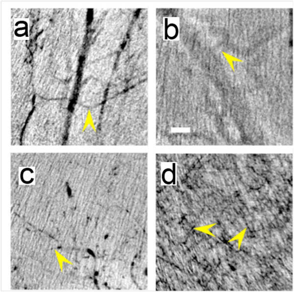
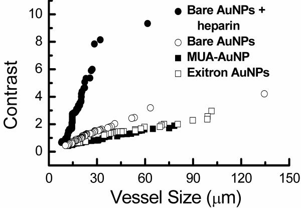
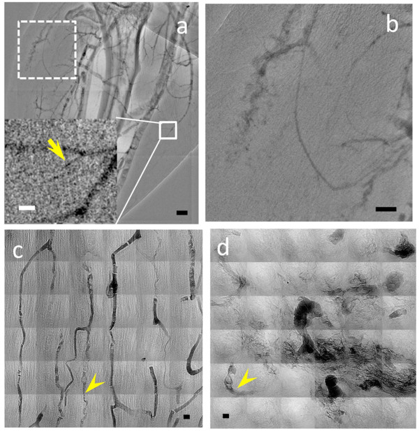
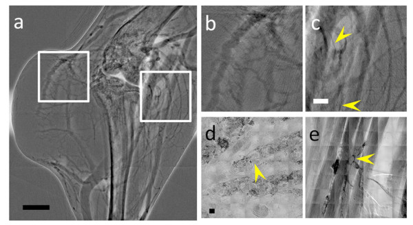
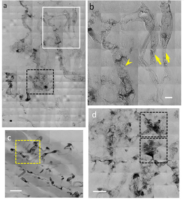
We tested different contrast agents based on gold nanoparticles (AuNPs) for the detection of cancer-related angiogenesis by synchrotron microradiology, microtomography and high resolution X-ray microscopy. Among them only bare-AuNPs in conjunction with heparin injection provided sufficient contrast to allow in vivo detection of small capillary species (the smallest measured lumen diameters were 3-5 μm). The detected vessel density was 3-7 times higher than with other nanoparticles. We also found that bare-AuNPs with heparin allows detecting symptoms of local extravascular nanoparticle diffusion in tumor areas where capillary leakage appeared.
Although high-Z AuNPs are natural candidates as radiology contrast agents, their success is not guaranteed, in particular when targeting very small blood vessels in tumor-related angiography. We found that AuNPs injected with heparin produced the contrast level needed to reveal--for the first time by X-ray imaging--tumor microvessels with 3-5 μm diameter as well as extravascular diffusion due to basal membrane defenestration. These results open the interesting possibility of functional imaging of the tumor microvasculature, of its development and organization, as well as of the effects of anti-angiogenic drugs.
血管生成与癌症的发展密切相关,特别是因为有可能进行早期检测和新的治疗策略。然而,由于成像技术在检测在血管生成应激下生长的微小血管(直径 3-5μm)方面的局限性,这些研究受到了负面影响。我们报告说,具有非常高空间分辨率的基于同步加速器的 X 射线成像技术可以克服这一障碍,只要使用合适的对比剂即可。
我们测试了不同的基于金纳米粒子(AuNPs)的对比剂,用于通过同步加速器微辐射学、微断层扫描和高分辨率 X 射线显微镜检测与癌症相关的血管生成。其中,只有裸露的 AuNPs 与肝素注射结合使用才能提供足够的对比度,从而能够在体内检测到小毛细血管(测量到的最小管腔直径为 3-5μm)。检测到的血管密度比其他纳米粒子高 3-7 倍。我们还发现,裸露的 AuNPs 与肝素一起使用可以检测到肿瘤区域中血管外纳米粒子扩散的局部症状,在那里毛细血管渗漏出现。
尽管高 Z 值的 AuNPs 是天然的放射学对比剂候选物,但它们的成功并不能保证,特别是在针对肿瘤相关血管造影中的非常小的血管时。我们发现,注射肝素的 AuNPs 产生了所需的对比度,可以首次通过 X 射线成像揭示直径为 3-5μm 的肿瘤微血管以及由于基底膜开窗引起的血管外扩散。这些结果为肿瘤微血管的功能成像、其发育和组织以及抗血管生成药物的效果开辟了有趣的可能性。