• 文献检索
  • 文档翻译
  • 深度研究
  • 学术资讯
  • Suppr Zotero 插件Zotero 插件
  • 邀请有礼
  • 套餐&价格
  • 历史记录
应用&插件
Suppr Zotero 插件Zotero 插件浏览器插件Mac 客户端Windows 客户端微信小程序
定价
高级版会员购买积分包购买API积分包
服务
文献检索文档翻译深度研究API 文档MCP 服务
关于我们
关于 Suppr公司介绍联系我们用户协议隐私条款
关注我们

Suppr 超能文献

核心技术专利:CN118964589B侵权必究
粤ICP备2023148730 号-1Suppr @ 2026

文献检索

告别复杂PubMed语法,用中文像聊天一样搜索,搜遍4000万医学文献。AI智能推荐,让科研检索更轻松。

立即免费搜索

文件翻译

保留排版,准确专业,支持PDF/Word/PPT等文件格式,支持 12+语言互译。

免费翻译文档

深度研究

AI帮你快速写综述,25分钟生成高质量综述,智能提取关键信息,辅助科研写作。

立即免费体验

糖尿病视网膜病变中的局部血视网膜屏障渗漏与视网膜光敏感性

Localised blood-retinal barrier leakage and retinal light sensitivity in diabetic retinopathy.

作者信息

Bek T, Lund-Andersen H

机构信息

Department of Ophthalmology, University of Copenhagen, Gentofte Hospital, Hellerup, Denmark.

出版信息

Br J Ophthalmol. 1990 Jul;74(7):388-92. doi: 10.1136/bjo.74.7.388.

DOI:10.1136/bjo.74.7.388
PMID:2378853
原文链接:https://pmc.ncbi.nlm.nih.gov/articles/PMC1042146/
Abstract

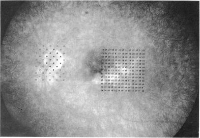
Twenty patients with insulin dependent diabetes mellitus were selected on the basis of morphological signs of blood-retinal barrier leakage--namely, hard exudates seen on fundus photographs and/or localised leakage of fluorescein seen on fluorescein angiograms. Computerised perimetry was carried out in visual field areas that corresponded to the morphological lesions, and the visual field data were accurately correlated with the morphology as seen on fundus photographs and fluorescein angiograms. In addition, in seven of the patients who represented the range of leakage among the patients studied, the blood-retinal barrier leakage was quantitated by vitreous fluorophotometry. In 16 cases normal light sensitivity was found in retinal areas showing localised leakage as studied on fluorescein angiograms. In four cases with pronounced maculopathy, where scotomata occurred, there was no topographical correlation between the scotomata and barrier leakage. Furthermore hard exudates often, but not consistently, caused localised scotomata when arranged in dense conglomerates. The permeability values correlated with angiographically observed hyperfluorescence in the macular area. On the basis of the techniques employed in the present study it seems that breakdown of the blood-retinal barrier is an earlier event than disturbance of neurosensory function in the development of diabetic retinopathy. However, the findings give no evidence of a causal relationship between barrier leakage and damage to sensory cell function.

摘要

根据血视网膜屏障渗漏的形态学体征,即眼底照片上可见的硬性渗出物和/或荧光素血管造影上可见的荧光素局部渗漏,选取了20例胰岛素依赖型糖尿病患者。在与形态学病变相对应的视野区域进行计算机视野检查,并将视野数据与眼底照片和荧光素血管造影上所见的形态学准确关联。此外,在代表所研究患者渗漏范围的7例患者中,通过玻璃体荧光光度法对血视网膜屏障渗漏进行了定量。在16例病例中,荧光素血管造影研究显示,视网膜局部渗漏区域的光敏感度正常。在4例有明显黄斑病变且出现暗点的病例中,暗点与屏障渗漏之间不存在地形学相关性。此外,硬性渗出物在密集聚集时,常但并非总是会导致局部暗点。通透性值与黄斑区血管造影观察到的高荧光相关。根据本研究采用的技术,在糖尿病视网膜病变的发展过程中,血视网膜屏障的破坏似乎比神经感觉功能障碍更早发生。然而,这些发现没有证据表明屏障渗漏与感觉细胞功能损害之间存在因果关系。

https://cdn.ncbi.nlm.nih.gov/pmc/blobs/704a/1042146/7f5aa49dfae5/brjopthal00581-0008-b.jpg
https://cdn.ncbi.nlm.nih.gov/pmc/blobs/704a/1042146/6956c1381438/brjopthal00581-0005-a.jpg
https://cdn.ncbi.nlm.nih.gov/pmc/blobs/704a/1042146/5bd58f950c5a/brjopthal00581-0006-a.jpg
https://cdn.ncbi.nlm.nih.gov/pmc/blobs/704a/1042146/2dd99fcdb868/brjopthal00581-0007-a.jpg
https://cdn.ncbi.nlm.nih.gov/pmc/blobs/704a/1042146/082ae8e39c05/brjopthal00581-0007-b.jpg
https://cdn.ncbi.nlm.nih.gov/pmc/blobs/704a/1042146/1ce8c3d458c7/brjopthal00581-0008-a.jpg
https://cdn.ncbi.nlm.nih.gov/pmc/blobs/704a/1042146/7f5aa49dfae5/brjopthal00581-0008-b.jpg

相似文献

1
Localised blood-retinal barrier leakage and retinal light sensitivity in diabetic retinopathy.糖尿病视网膜病变中的局部血视网膜屏障渗漏与视网膜光敏感性
Br J Ophthalmol. 1990 Jul;74(7):388-92. doi: 10.1136/bjo.74.7.388.
2
Localized retinal morphology and differential light sensitivity in diabetic retinopathy. Methodology and clinical results.糖尿病视网膜病变中的局部视网膜形态和差异光敏感性。方法与临床结果。
Acta Ophthalmol Suppl (1985). 1992(207):1-36.
3
Localised scotomata and types of vascular occlusion in diabetic retinopathy.糖尿病视网膜病变中的局限性暗点及血管阻塞类型
Acta Ophthalmol (Copenh). 1991 Feb;69(1):11-8. doi: 10.1111/j.1755-3768.1991.tb01984.x.
4
Three-year follow-up study of blood-retinal barrier and retinal thickness alterations in patients with type 2 diabetes mellitus and mild nonproliferative diabetic retinopathy.2型糖尿病合并轻度非增殖性糖尿病视网膜病变患者血视网膜屏障及视网膜厚度改变的三年随访研究
Arch Ophthalmol. 2004 Feb;122(2):211-7. doi: 10.1001/archopht.122.2.211.
5
Cotton-wool spots and retinal light sensitivity in diabetic retinopathy.糖尿病视网膜病变中的棉絮斑与视网膜光敏感度
Br J Ophthalmol. 1991 Jan;75(1):13-7. doi: 10.1136/bjo.75.1.13.
6
Alterations of the blood-retinal barrier and retinal thickness in preclinical retinopathy in subjects with type 2 diabetes.2型糖尿病患者临床前期视网膜病变时血视网膜屏障和视网膜厚度的改变。
Arch Ophthalmol. 2000 Oct;118(10):1364-9. doi: 10.1001/archopht.118.10.1364.
7
Effect of morphological abnormalities on blood retinal barrier permeability in diabetic retinopathy.形态学异常对糖尿病视网膜病变中血视网膜屏障通透性的影响。
Doc Ophthalmol. 1992;80(2):183-8. doi: 10.1007/BF00161244.
8
One-year follow-up of blood-retinal barrier and retinal thickness alterations in patients with type 2 diabetes mellitus and mild nonproliferative retinopathy.2型糖尿病合并轻度非增殖性视网膜病变患者血视网膜屏障及视网膜厚度改变的一年随访
Arch Ophthalmol. 2001 Oct;119(10):1469-74. doi: 10.1001/archopht.119.10.1469.
9
Coexistence of localized scotomata and neovascularizations in proliferative diabetic retinopathy.增殖性糖尿病视网膜病变中局部性暗点与新生血管的共存。
Acta Ophthalmol (Copenh). 1990 Aug;68(4):421-7. doi: 10.1111/j.1755-3768.1990.tb01670.x.
10
Mapping retinal fluorescein leakage with confocal scanning laser fluorometry of the human vitreous.通过共焦扫描激光荧光测定法绘制人玻璃体视网膜荧光素渗漏情况。
Arch Ophthalmol. 1999 May;117(5):631-7. doi: 10.1001/archopht.117.5.631.

引用本文的文献

1
Diabetic vascular hyperpermeability: optical coherence tomography angiography and functional loss assessments of relationships among retinal vasculature changes.糖尿病血管通透性增加:视网膜血管变化与功能丧失之间关系的光相干断层扫描血管造影和评估。
Sci Rep. 2021 Feb 18;11(1):4185. doi: 10.1038/s41598-021-83334-6.
2
Plasma metabolomic profiling of proliferative diabetic retinopathy.增殖性糖尿病视网膜病变的血浆代谢组学分析
Nutr Metab (Lond). 2019 May 28;16:37. doi: 10.1186/s12986-019-0358-3. eCollection 2019.
3
Visualizing Structure and Vascular Interactions: Macular Nonperfusion in Three Capillary Plexuses.

本文引用的文献

1
Quantitative vitreous fluorophotometry applying a mathematical model of the eye.应用眼部数学模型的定量玻璃体荧光光度测定法。
Invest Ophthalmol Vis Sci. 1985 May;26(5):698-710.
2
Diabetic retinopathy viewed as a neurosensory disorder.
Arch Ophthalmol. 1986 Jul;104(7):989-90. doi: 10.1001/archopht.1986.01050190047037.
3
Vitreous fluorophotometry: mathematical analysis of the effect of peripheral leakage on axial scans.玻璃体荧光光度测定法:周边渗漏对轴向扫描影响的数学分析。
Invest Ophthalmol Vis Sci. 1989 Jul;30(7):1522-6.
可视化结构与血管相互作用:三个毛细血管丛中的黄斑无灌注
Ophthalmic Surg Lasers Imaging Retina. 2018 Nov 1;49(11):e182-e190. doi: 10.3928/23258160-20181101-16.
4
High-Resolution Imaging by Adaptive Optics Scanning Laser Ophthalmoscopy Reveals Two Morphologically Distinct Types of Retinal Hard Exudates.自适应光学扫描激光检眼镜的高分辨率成像显示两种形态不同的视网膜硬性渗出物。
Sci Rep. 2016 Sep 19;6:33574. doi: 10.1038/srep33574.
5
Retinal Sensitivity over Hard Exudates in Diabetic Retinopathy.糖尿病视网膜病变中硬性渗出物上方的视网膜敏感性
J Ophthalmic Vis Res. 2015 Apr-Jun;10(2):160-4. doi: 10.4103/2008-322X.163771.
6
Short-wavelength automated perimetry in patients with diabetes mellitus without macular edema.
Graefes Arch Clin Exp Ophthalmol. 2003 Jun;241(6):468-71. doi: 10.1007/s00417-003-0666-0. Epub 2003 May 17.
7
The relation between visual acuity, fixation stability, and the size and location of foveal hard exudates after photocoagulation for diabetic maculopathy: a 1-year follow-up study.
Graefes Arch Clin Exp Ophthalmol. 2003 Jun;241(6):458-62. doi: 10.1007/s00417-003-0661-5. Epub 2003 May 15.
8
Visual field loss after argon laser panretinal photocoagulation in diabetic retinopathy: full- versus mild-scatter coagulation.糖尿病性视网膜病变患者接受氩激光全视网膜光凝术后的视野缺损:全散射与轻度散射光凝的比较
Int Ophthalmol. 1998;22(5):313-9. doi: 10.1023/a:1006367029134.
9
Automated static perimetry as a screening method for evaluation of retinal perfusion in diabetic retinopathy.自动静态视野检查作为评估糖尿病视网膜病变视网膜灌注的筛查方法。
Int Ophthalmol. 1997;21(6):305-9. doi: 10.1023/a:1006024305992.
10
Effect of diabetes on levels and uptake of putative amino acid neurotransmitters in rat retina and retinal pigment epithelium.
Neurochem Res. 1996 Oct;21(10):1167-71. doi: 10.1007/BF02532391.
4
The influence of stimulus size on perimetric detection of small scotomata.刺激大小对小暗点视野检测的影响。
Graefes Arch Clin Exp Ophthalmol. 1989;227(6):531-4. doi: 10.1007/BF02169446.
5
Accurate superimposition of perimetry data onto fundus photographs.
Acta Ophthalmol (Copenh). 1990 Feb;68(1):11-8. doi: 10.1111/j.1755-3768.1990.tb01642.x.
6
Early breakdown of the blood-retinal barrier in diabetes.糖尿病中血视网膜屏障的早期破坏。
Br J Ophthalmol. 1975 Nov;59(11):649-56. doi: 10.1136/bjo.59.11.649.