• 文献检索
  • 文档翻译
  • 深度研究
  • 学术资讯
  • Suppr Zotero 插件Zotero 插件
  • 邀请有礼
  • 套餐&价格
  • 历史记录
应用&插件
Suppr Zotero 插件Zotero 插件浏览器插件Mac 客户端Windows 客户端微信小程序
定价
高级版会员购买积分包购买API积分包
服务
文献检索文档翻译深度研究API 文档MCP 服务
关于我们
关于 Suppr公司介绍联系我们用户协议隐私条款
关注我们

Suppr 超能文献

核心技术专利:CN118964589B侵权必究
粤ICP备2023148730 号-1Suppr @ 2026

文献检索

告别复杂PubMed语法,用中文像聊天一样搜索,搜遍4000万医学文献。AI智能推荐,让科研检索更轻松。

立即免费搜索

文件翻译

保留排版,准确专业,支持PDF/Word/PPT等文件格式,支持 12+语言互译。

免费翻译文档

深度研究

AI帮你快速写综述,25分钟生成高质量综述,智能提取关键信息,辅助科研写作。

立即免费体验

人体膝关节软骨-骨过渡结构的形态学特征及骨软骨支架的计算机辅助设计

Morphological characteristics of cartilage-bone transitional structures in the human knee joint and CAD design of an osteochondral scaffold.

作者信息

Bian Weiguo, Lian Qin, Li Dichen, Wang Jin, Zhang Weijie, Jin Zhongmin, Qiu Yusheng

机构信息

The First Affiliated Hospital of Xi'an Jiaotong University, Xi'an, 710061, Shaanxi, China.

State Key Lab for Manufacturing System Engineering, Xi'an Jiaotong University, Xi'an, 710049, Shaanxi, China.

出版信息

Biomed Eng Online. 2016 Jul 14;15(1):82. doi: 10.1186/s12938-016-0200-3.

DOI:10.1186/s12938-016-0200-3
PMID:27418247
原文链接:https://pmc.ncbi.nlm.nih.gov/articles/PMC4944264/
Abstract

BACKGROUND

There is a lack of understanding of the morphological characteristics of the cartilage-bone interface. Materials that are currently being used in tissue engineering do not adequately support the regeneration of bone and cartilage tissues. The present study aimed to explore the morphological characteristics of cartilage-bone transitional structures in the human knee joint and to design a biomimetic osteochondral scaffold based on morphological data.

METHODS

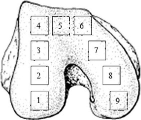
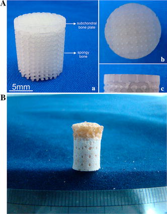
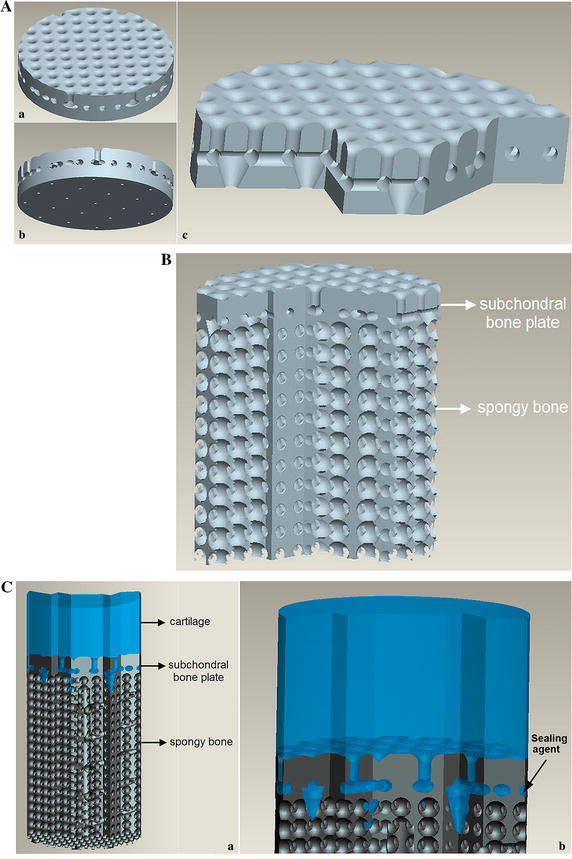
Histology, micro-computed tomography (micro-CT), and scanning electron microscopy (SEM) were used to investigate the microstructure of the cartilage-bone transitional structures. Morphological characteristics and their distribution were obtained and summarized into a biomimetic design. A three-dimensional model of a biomimetic osteochondral scaffold was CAD designed. A prototype of the resulting subchondral bone scaffold was constructed by stereolithography using resin.

RESULTS

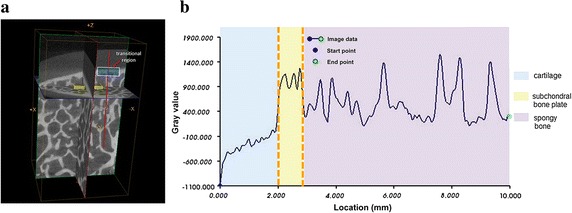
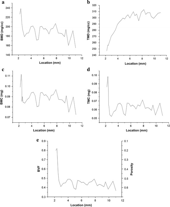
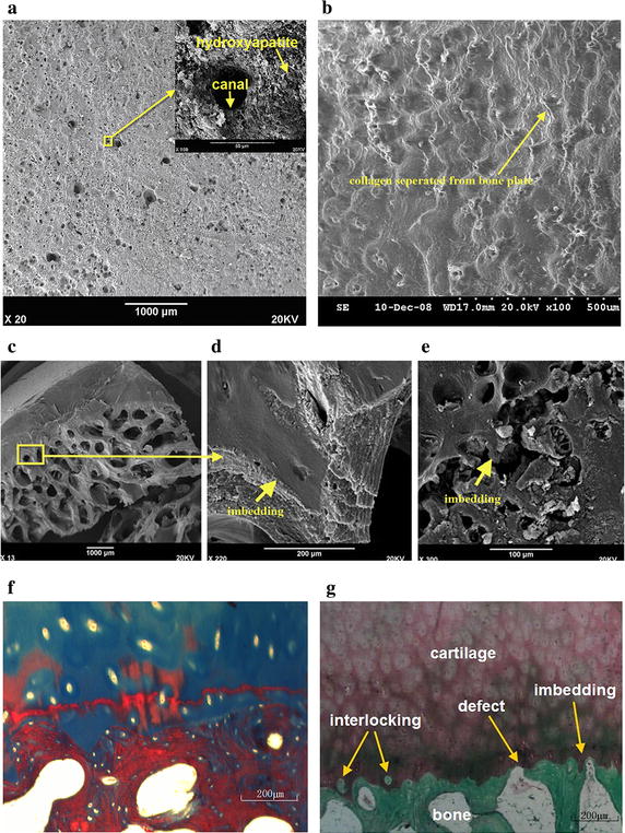
Micro-CT revealed that subchondral bone presented a gradually changing structure from the subchondral to spongy bone tissue. The subchondral bone plate was more compact with ~20 % porosity compared with ~60 % porosity for the spongy bone. Histology and SEM showed that cartilage was stabilized on the subchondral bone plate by conjunctions, imbedding, interlocking, and binding forces generated by collagen fibers. Some scattered defects allow blood vessel invasion and nutritional supply.

CONCLUSIONS

The subchondral bone plate is not an intact plate between the cartilage and bone cavity, and some scattered defects exist that allow blood vessel invasion and nutritional supply. This characteristic was used to design an osteochondral scaffold. This could be used to construct an osteochondral complex that is similar to native bones.

摘要

背景

目前对软骨-骨界面的形态学特征缺乏了解。组织工程中目前使用的材料不能充分支持骨和软骨组织的再生。本研究旨在探索人类膝关节软骨-骨过渡结构的形态学特征,并基于形态学数据设计一种仿生骨软骨支架。

方法

采用组织学、微计算机断层扫描(micro-CT)和扫描电子显微镜(SEM)研究软骨-骨过渡结构的微观结构。获取形态学特征及其分布并总结成仿生设计。利用计算机辅助设计(CAD)构建仿生骨软骨支架的三维模型。使用树脂通过立体光刻技术构建所得软骨下骨支架的原型。

结果

Micro-CT显示,软骨下骨呈现出从软骨下到松质骨组织逐渐变化的结构。软骨下骨板更致密,孔隙率约为20%,而松质骨的孔隙率约为60%。组织学和SEM显示,软骨通过胶原纤维产生的连接、嵌入、互锁和结合力稳定在软骨下骨板上。一些分散的缺陷允许血管侵入和营养供应。

结论

软骨下骨板不是软骨和骨腔之间的完整板,存在一些分散的缺陷,允许血管侵入和营养供应。利用这一特性设计了一种骨软骨支架。这可用于构建类似于天然骨的骨软骨复合体。

https://cdn.ncbi.nlm.nih.gov/pmc/blobs/beb7/4944264/d928421ddb7b/12938_2016_200_Fig6_HTML.jpg
https://cdn.ncbi.nlm.nih.gov/pmc/blobs/beb7/4944264/beb80ac4ed43/12938_2016_200_Fig1_HTML.jpg
https://cdn.ncbi.nlm.nih.gov/pmc/blobs/beb7/4944264/f1433d589649/12938_2016_200_Fig2_HTML.jpg
https://cdn.ncbi.nlm.nih.gov/pmc/blobs/beb7/4944264/eebb2577e358/12938_2016_200_Fig3_HTML.jpg
https://cdn.ncbi.nlm.nih.gov/pmc/blobs/beb7/4944264/53e08492fded/12938_2016_200_Fig4_HTML.jpg
https://cdn.ncbi.nlm.nih.gov/pmc/blobs/beb7/4944264/134dbbabef8e/12938_2016_200_Fig5_HTML.jpg
https://cdn.ncbi.nlm.nih.gov/pmc/blobs/beb7/4944264/d928421ddb7b/12938_2016_200_Fig6_HTML.jpg

相似文献

1
Morphological characteristics of cartilage-bone transitional structures in the human knee joint and CAD design of an osteochondral scaffold.人体膝关节软骨-骨过渡结构的形态学特征及骨软骨支架的计算机辅助设计
Biomed Eng Online. 2016 Jul 14;15(1):82. doi: 10.1186/s12938-016-0200-3.
2
3D-printed scaffolds with calcified layer for osteochondral tissue engineering.用于骨软骨组织工程的带钙化层的3D打印支架。
J Biosci Bioeng. 2018 Sep;126(3):389-396. doi: 10.1016/j.jbiosc.2018.03.014. Epub 2018 May 16.
3
Biomimetic design and fabrication of multilayered osteochondral scaffolds by low-temperature deposition manufacturing and thermal-induced phase-separation techniques.通过低温沉积制造和热诱导相分离技术进行多层骨软骨支架的仿生设计与制造。
Biofabrication. 2017 May 23;9(2):025021. doi: 10.1088/1758-5090/aa7078.
4
[Cartilage repair and subchondral bone reconstruction based on three-dimensional printing technique].基于三维打印技术的软骨修复与软骨下骨重建
Zhongguo Xiu Fu Chong Jian Wai Ke Za Zhi. 2014 Mar;28(3):318-24.
5
Small subchondral drill holes improve marrow stimulation of articular cartilage defects.小的软骨下钻孔可改善关节软骨缺损的骨髓刺激。
Am J Sports Med. 2014 Nov;42(11):2741-50. doi: 10.1177/0363546514547029. Epub 2014 Aug 28.
6
A single integrated osteochondral in situ composite scaffold with a multi-layered functional structure.具有多层次功能结构的一体化原位复合骨软骨支架
Colloids Surf B Biointerfaces. 2018 Jul 1;167:354-363. doi: 10.1016/j.colsurfb.2018.04.029. Epub 2018 Apr 16.
7
Evaluation of an extracellular matrix-derived acellular biphasic scaffold/cell construct in the repair of a large articular high-load-bearing osteochondral defect in a canine model.评价细胞外基质衍生去细胞双相支架/细胞构建体在犬模型中大关节高负荷承重骨软骨缺损修复中的作用。
Chin Med J (Engl). 2011 Dec;124(23):3930-8.
8
Design and characterization of a tissue-engineered bilayer scaffold for osteochondral tissue repair.用于骨软骨组织修复的组织工程双层支架的设计与表征
J Tissue Eng Regen Med. 2015 Oct;9(10):1182-92. doi: 10.1002/term.1651. Epub 2012 Nov 21.
9
Autologous tissue transplantations for osteochondral repair.用于骨软骨修复的自体组织移植
Dan Med J. 2016 Apr;63(4).
10
Recent Approaches to the Manufacturing of Biomimetic Multi-Phasic Scaffolds for Osteochondral Regeneration.用于骨软骨再生的仿生多相支架制造的最新方法。
Int J Mol Sci. 2018 Jun 13;19(6):1755. doi: 10.3390/ijms19061755.

引用本文的文献

1
3D Printing of Bone Substitutes Based on Vat Photopolymerization Processes: A Systematic Review.基于光固化成型工艺的骨替代物3D打印:一项系统综述
J Tissue Eng Regen Med. 2023 Apr 8;2023:3901448. doi: 10.1155/2023/3901448. eCollection 2023.
2
Effect of collagen-based scaffolds with hydroxyapatite on the repair of cartilage defects in the rabbit knee joint.含羟基磷灰石的胶原基支架对兔膝关节软骨缺损修复的影响。
J Orthop Surg Res. 2024 Dec 3;19(1):818. doi: 10.1186/s13018-024-05323-5.
3
3D printing tissue-engineered scaffolds for auricular reconstruction.

本文引用的文献

1
Bone mineral density in cone beam computed tomography: Only a few shades of gray.锥形束计算机断层扫描中的骨密度:仅有几种灰度。
World J Radiol. 2014 Aug 28;6(8):607-12. doi: 10.4329/wjr.v6.i8.607.
2
Metabolic profiling of isomeric aglycones central-icaritin (c-IT) and icaritin (IT) in osteoporotic rats by UPLC-QTOF-MS.采用超高效液相色谱-四极杆飞行时间质谱法对骨质疏松大鼠体内异构体苷元中-淫羊藿素(c-IT)和淫羊藿素(IT)进行代谢谱分析。
Drug Test Anal. 2015 Apr;7(4):309-19. doi: 10.1002/dta.1672. Epub 2014 Jun 16.
3
Osteochondral scaffold reconstruction for complex knee lesions: a comparative evaluation.
用于耳廓重建的3D打印组织工程支架
Mater Today Bio. 2024 Jul 2;27:101141. doi: 10.1016/j.mtbio.2024.101141. eCollection 2024 Aug.
4
Knee Measurement System with Osteoarthritis Levels Using Artificial Cartilage and Skeletons.使用人工软骨和骨骼的骨关节炎分级膝关节测量系统。
Biomimetics (Basel). 2024 Mar 8;9(3):166. doi: 10.3390/biomimetics9030166.
5
Microarchitecture Alternations of Osteochondral Junction in Patients with Osteonecrosis of the Femoral Head.股骨头坏死患者的软骨-骨连接的微观结构改变。
Calcif Tissue Int. 2024 Feb;114(2):119-128. doi: 10.1007/s00223-023-01153-5. Epub 2023 Nov 30.
6
Intra-osseous plasma rich in growth factors enhances cartilage and subchondral bone regeneration in rabbits with acute full thickness chondral defects: Histological assessment.富含生长因子的骨内血浆可促进急性全层软骨缺损兔的软骨和软骨下骨再生:组织学评估
Front Vet Sci. 2023 Mar 29;10:1131666. doi: 10.3389/fvets.2023.1131666. eCollection 2023.
7
Acrylamide-based hydrogels with distinct osteogenic and chondrogenic differentiation potential.具有不同成骨和成软骨分化潜能的基于丙烯酰胺的水凝胶。
Prog Biomater. 2022 Sep;11(3):297-309. doi: 10.1007/s40204-022-00196-5. Epub 2022 Jul 16.
8
Biomimetic Approaches for the Design and Fabrication of Bone-to-Soft Tissue Interfaces.仿生学方法在骨-软组织界面设计与制造中的应用。
ACS Biomater Sci Eng. 2023 Jul 10;9(7):3810-3831. doi: 10.1021/acsbiomaterials.1c00620. Epub 2021 Nov 16.
9
Comparison of chondrogenesis-related biological behaviors between human urine-derived stem cells and human bone marrow mesenchymal stem cells from the same individual.比较人尿液来源干细胞与同个体来源骨髓间充质干细胞的软骨生成相关生物学行为。
Stem Cell Res Ther. 2021 Jun 28;12(1):366. doi: 10.1186/s13287-021-02370-1.
10
Subchondral Bone Remodeling: A Therapeutic Target for Osteoarthritis.软骨下骨重塑:骨关节炎的一个治疗靶点。
Front Cell Dev Biol. 2021 Jan 21;8:607764. doi: 10.3389/fcell.2020.607764. eCollection 2020.
复杂膝关节损伤的骨软骨支架重建:一项对比评估
Knee. 2013 Dec;20(6):570-6. doi: 10.1016/j.knee.2013.05.007. Epub 2013 Jun 28.
4
Optimization of composition, structure and mechanical strength of bioactive 3-D glass-ceramic scaffolds for bone substitution.用于骨替代的生物活性 3-D 玻璃陶瓷支架的组成、结构和机械强度的优化。
J Biomater Appl. 2013 Mar;27(7):872-90. doi: 10.1177/0885328211429193. Epub 2011 Dec 29.
5
A pilot study of the use of an osteochondral scaffold plug for cartilage repair in the knee and how to deal with early clinical failures.一项关于使用骨软骨支架塞进行膝关节软骨修复的初步研究,以及如何应对早期临床失败。
Arthroscopy. 2012 Feb;28(2):225-33. doi: 10.1016/j.arthro.2011.07.017. Epub 2011 Oct 20.
6
The effects of high-dose, long-term alendronate treatment on microarchitecture and bone mineral density of compact and trabecular bone in the proximal femur of adult male rabbits.大剂量、长期阿伦膦酸钠治疗对成年雄性兔股骨近端密质骨和松质骨微观结构和骨密度的影响。
Arch Orthop Trauma Surg. 2010 Jul;130(7):937-44. doi: 10.1007/s00402-010-1116-1. Epub 2010 May 20.
7
Histomorphometric analysis of adult articular calcified cartilage zone.成人关节钙化软骨带的组织形态计量分析。
J Struct Biol. 2009 Dec;168(3):359-65. doi: 10.1016/j.jsb.2009.08.010. Epub 2009 Aug 31.
8
Design of graded biomimetic osteochondral composite scaffolds.梯度仿生骨软骨复合支架的设计
Biomaterials. 2008 Sep;29(26):3539-46. doi: 10.1016/j.biomaterials.2008.05.008. Epub 2008 Jun 5.
9
Engineering custom-designed osteochondral tissue grafts.工程定制化骨软骨组织移植物。
Trends Biotechnol. 2008 Apr;26(4):181-9. doi: 10.1016/j.tibtech.2007.12.009. Epub 2008 Mar 4.
10
Cartilage damage pattern in relation to subchondral plate thickness in a collagenase-induced model of osteoarthritis.胶原酶诱导的骨关节炎模型中软骨损伤模式与软骨下骨板厚度的关系
Osteoarthritis Cartilage. 2008 Apr;16(4):506-14. doi: 10.1016/j.joca.2007.08.005. Epub 2007 Sep 27.