Pisciotta John M, Scholl Peter F, Shuman Joel L, Shualev Vladimir, Sullivan David J
Department of Molecular Microbiology and Immunology, Johns Hopkins University, Bloomberg School of Public Health, 615 N. Wolfe St., Baltimore, MD 21205-2179, USA.
Department of Environmental Health Sciences, Bloomberg School of Public Health, Johns Hopkins University, Baltimore, MD 21205-2103, USA.
Int J Parasitol Drugs Drug Resist. 2017 Apr;7(1):110-119. doi: 10.1016/j.ijpddr.2017.02.001. Epub 2017 Feb 8.
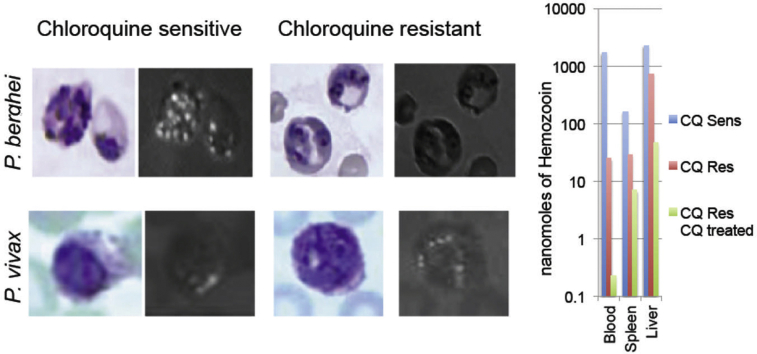
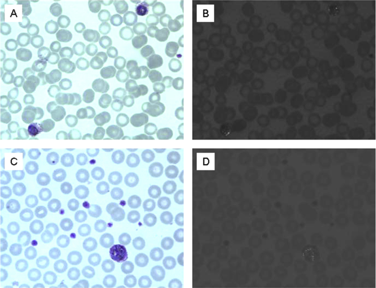
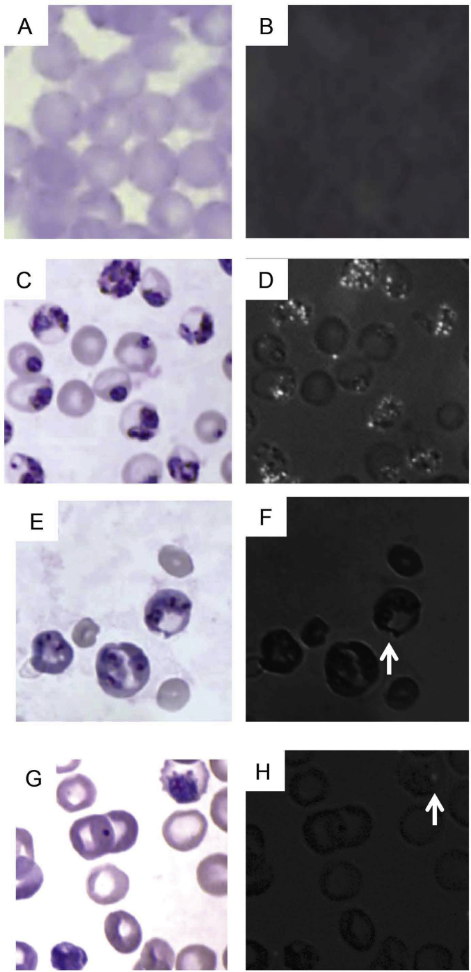
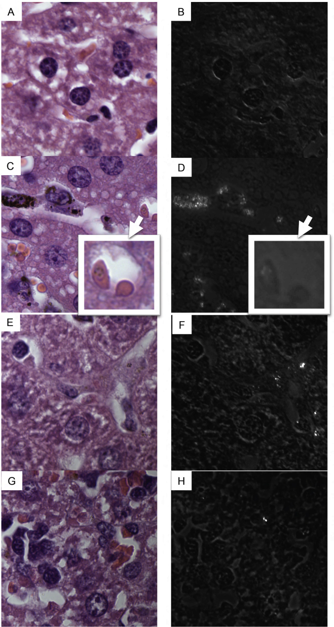
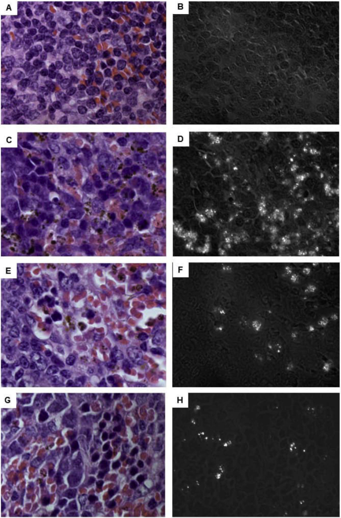
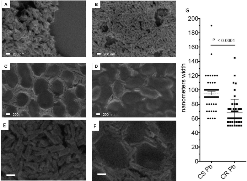
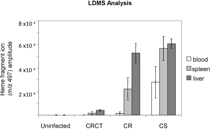
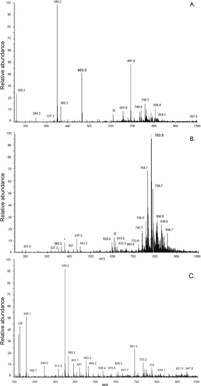
The incidence and global distribution of chloroquine resistant (CR) Plasmodium vivax infection has increased since emerging in 1989. The mechanism of resistance in CR P. vivax has not been defined. The resistance likely relates to the formation and disposition of hemozoin as chloroquine's primary mechanism of action involves disruption of hemozoin formation. CR P. berghei strains, like CR P. vivax strains, are confined to reticulocyte host cells and reportedly they do not accumulate appreciable intraerythrocytic hemozoin. Reports comparing hemozoin production between P. vivax strains and CR to chloroquine sensitive (CS) P. berghei are absent. Here we compare in vivo patterns of hemozoin formation and distribution in blood, spleen and liver tissue of male Swiss mice infected with CS or CR P. berghei not treated with chloroquine and CR P. berghei also treated with chloroquine. Light microscopy, laser desorption mass spectrometry and a colorimetric hemozoin assay detect trace hemozoin in the blood of CR P. berghei infected mice but significant hemozoin accumulation in liver and spleen tissue. Field emission in lens scanning electron microscopy reveals CR P. berghei hemozoin crystals are morphologically smaller but similar to those formed by CS parasites. CR P. berghei produces approximately five-fold less total hemozoin than CS strain. Lipid analysis of CS and CR P. berghei sucrose gradient purified bloodstage hemozoin indicates a similar lipid environment around the isolated hemozoin, predominately monopalmitic glycerol and monostearic glycerol. In contrast to CR and CS P. berghei, colorimetric hemozoin analysis of P. vivax strains indicates similar amounts of hemozoin are produced despite differing chloroquine sensitivities. These results suggest CR P. berghei forms significant hemozoin which accumulates in liver and spleen tissues and that the P. vivax chloroquine resistance mechanism differs from P. berghei.
自1989年出现以来,氯喹耐药间日疟原虫感染的发病率及其在全球的分布有所增加。氯喹耐药间日疟原虫的耐药机制尚未明确。由于氯喹的主要作用机制涉及干扰疟原虫疟色素的形成,因此这种耐药性可能与疟色素的形成和分布有关。氯喹耐药伯氏疟原虫菌株与氯喹耐药间日疟原虫菌株一样,局限于网织红细胞宿主细胞,据报道它们不会在红细胞内积累可观的疟色素。目前尚无比较间日疟原虫菌株与氯喹敏感伯氏疟原虫疟色素产生情况的报告。在此,我们比较了未用氯喹治疗的氯喹敏感或氯喹耐药伯氏疟原虫以及用氯喹治疗的氯喹耐药伯氏疟原虫感染的雄性瑞士小鼠血液、脾脏和肝脏组织中疟色素形成和分布的体内模式。光学显微镜、激光解吸质谱法和比色法疟色素测定法检测到氯喹耐药伯氏疟原虫感染小鼠血液中有微量疟色素,但在肝脏和脾脏组织中有大量疟色素积累。透镜场发射扫描电子显微镜显示,氯喹耐药伯氏疟原虫的疟色素晶体在形态上较小,但与氯喹敏感寄生虫形成的晶体相似。氯喹耐药伯氏疟原虫产生的总疟色素比氯喹敏感菌株少约五倍。对氯喹敏感和氯喹耐药伯氏疟原虫经蔗糖梯度纯化的血期疟色素进行脂质分析表明,分离出的疟色素周围的脂质环境相似,主要是单棕榈酸甘油和单硬脂酸甘油。与氯喹耐药和氯喹敏感伯氏疟原虫不同,间日疟原虫菌株的比色法疟色素分析表明,尽管氯喹敏感性不同,但产生的疟色素量相似。这些结果表明,氯喹耐药伯氏疟原虫形成大量疟色素并在肝脏和脾脏组织中积累,且间日疟原虫的氯喹耐药机制与伯氏疟原虫不同。