• 文献检索
  • 文档翻译
  • 深度研究
  • 学术资讯
  • Suppr Zotero 插件Zotero 插件
  • 邀请有礼
  • 套餐&价格
  • 历史记录
应用&插件
Suppr Zotero 插件Zotero 插件浏览器插件Mac 客户端Windows 客户端微信小程序
定价
高级版会员购买积分包购买API积分包
服务
文献检索文档翻译深度研究API 文档MCP 服务
关于我们
关于 Suppr公司介绍联系我们用户协议隐私条款
关注我们

Suppr 超能文献

核心技术专利:CN118964589B侵权必究
粤ICP备2023148730 号-1Suppr @ 2026

文献检索

告别复杂PubMed语法,用中文像聊天一样搜索,搜遍4000万医学文献。AI智能推荐,让科研检索更轻松。

立即免费搜索

文件翻译

保留排版,准确专业,支持PDF/Word/PPT等文件格式,支持 12+语言互译。

免费翻译文档

深度研究

AI帮你快速写综述,25分钟生成高质量综述,智能提取关键信息,辅助科研写作。

立即免费体验

通过数字全息显微镜数据的图像处理评估恶性细胞的转移潜能。

Evaluation of the metastatic potential of malignant cells by image processing of digital holographic microscopy data.

作者信息

Calin Violeta L, Mihailescu Mona, Scarlat Eugen I, Baluta Alexandra V, Calin Daniel, Kovacs Eugenia, Savopol Tudor, Moisescu Mihaela G

机构信息

Biophysics and Cellular Biotechnology Department Faculty of Medicine Carol Davila University of Medicine and Pharmacy Bucharest Romania.

Physics Department Faculty of Applied Sciences Politehnica University of Bucharest Romania.

出版信息

FEBS Open Bio. 2017 Sep 2;7(10):1527-1538. doi: 10.1002/2211-5463.12282. eCollection 2017 Oct.

DOI:10.1002/2211-5463.12282
PMID:28979841
原文链接:https://pmc.ncbi.nlm.nih.gov/articles/PMC5623698/
Abstract

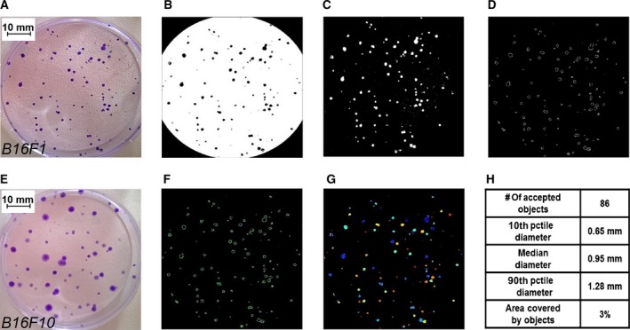
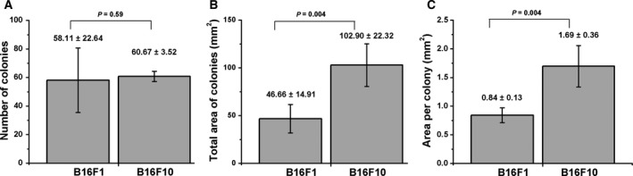
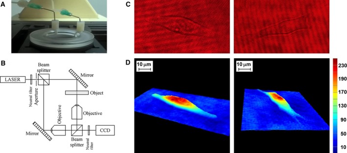
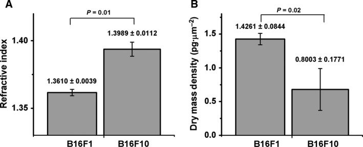
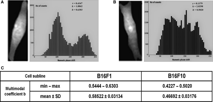
The cell refractive index has been proposed as a putative cancer biomarker of great potential, being correlated with cell content and morphology, cell division rate and membrane permeability. We used digital holographic microscopy to compare the refractive index and dry mass density of two B16 murine melanoma sublines of different metastatic potential. Using statistical methods, the distribution of phase shifts within the reconstructed quantitative phase images was analyzed by the method of bimodality coefficients. The observed correlation of refractive index, dry mass density and bimodality profile with the metastatic potential of the cells was validated by real time impedance-based assay and clonogenic tests. We suggest that the refractive index and bimodality analysis of quantitative phase image histograms could be developed as optical biomarkers useful in label-free detection and quantitative evaluation of cell metastatic potential.

摘要

细胞折射率已被认为是一种极具潜力的假定癌症生物标志物,它与细胞内容物和形态、细胞分裂速率及膜通透性相关。我们使用数字全息显微镜比较了两种具有不同转移潜能的B16小鼠黑色素瘤亚系的折射率和干质量密度。通过统计方法,采用双峰系数法分析了重建的定量相位图像内的相移分布。通过基于实时阻抗的检测和克隆形成试验验证了所观察到的折射率、干质量密度和双峰分布与细胞转移潜能之间的相关性。我们认为,定量相位图像直方图的折射率和双峰分析可发展成为用于无标记检测和细胞转移潜能定量评估的光学生物标志物。

https://cdn.ncbi.nlm.nih.gov/pmc/blobs/26d2/5623698/6cac89d90d33/FEB4-7-1527-g006.jpg
https://cdn.ncbi.nlm.nih.gov/pmc/blobs/26d2/5623698/e33be3c41d8f/FEB4-7-1527-g001.jpg
https://cdn.ncbi.nlm.nih.gov/pmc/blobs/26d2/5623698/a674d355a348/FEB4-7-1527-g002.jpg
https://cdn.ncbi.nlm.nih.gov/pmc/blobs/26d2/5623698/860acdd9a5b7/FEB4-7-1527-g003.jpg
https://cdn.ncbi.nlm.nih.gov/pmc/blobs/26d2/5623698/65cdf39eedc8/FEB4-7-1527-g004.jpg
https://cdn.ncbi.nlm.nih.gov/pmc/blobs/26d2/5623698/d75ccd93646d/FEB4-7-1527-g005.jpg
https://cdn.ncbi.nlm.nih.gov/pmc/blobs/26d2/5623698/6cac89d90d33/FEB4-7-1527-g006.jpg

相似文献

1
Evaluation of the metastatic potential of malignant cells by image processing of digital holographic microscopy data.通过数字全息显微镜数据的图像处理评估恶性细胞的转移潜能。
FEBS Open Bio. 2017 Sep 2;7(10):1527-1538. doi: 10.1002/2211-5463.12282. eCollection 2017 Oct.
2
Measurement of morphology thickness and refractive index in a melanoma A375 cell line using digital holographic microscopy.使用数字全息显微镜测量黑素瘤 A375 细胞系的形态厚度和折射率。
Appl Opt. 2021 Feb 1;60(4):815-822. doi: 10.1364/AO.410993.
3
A practical criterion for focusing of unstained cell samples using a digital holographic microscope.使用数字全息显微镜聚焦未染色细胞样品的实用标准。
J Microsc. 2020 Aug;279(2):114-122. doi: 10.1111/jmi.12924. Epub 2020 Jun 5.
4
Quantitative phase imaging of cells in a flow cytometry arrangement utilizing Michelson interferometer-based off-axis digital holographic microscopy.利用基于迈克尔逊干涉仪的离轴数字全息显微镜在流式细胞仪配置中对细胞进行定量相成像。
J Biophotonics. 2019 Sep;12(9):e201900085. doi: 10.1002/jbio.201900085. Epub 2019 Jun 20.
5
Integral refractive index determination of living suspension cells by multifocus digital holographic phase contrast microscopy.利用多焦点数字全息相衬显微镜测定活悬浮细胞的积分折射率
J Biomed Opt. 2007 Sep-Oct;12(5):054009. doi: 10.1117/1.2798639.
6
Digital holographic microscopy: a quantitative label-free microscopy technique for phenotypic screening.数字全息显微镜:一种用于表型筛选的无标记定量显微镜技术。
Comb Chem High Throughput Screen. 2014 Jan;17(1):80-8. doi: 10.2174/13862073113166660062.
7
Digital holographic microscopy evaluation of dynamic cell response to electroporation.数字全息显微镜对细胞电穿孔动态响应的评估
Biomed Opt Express. 2021 Mar 31;12(4):2519-2530. doi: 10.1364/BOE.421959. eCollection 2021 Apr 1.
8
Comparative study of human erythrocytes by digital holographic microscopy, confocal microscopy, and impedance volume analyzer.利用数字全息显微镜、共聚焦显微镜和阻抗体积分析仪对人类红细胞进行的对比研究。
Cytometry A. 2008 Oct;73(10):895-903. doi: 10.1002/cyto.a.20605.
9
Cell refractive index tomography by digital holographic microscopy.基于数字全息显微镜的细胞折射率断层扫描
Opt Lett. 2006 Jan 15;31(2):178-80. doi: 10.1364/ol.31.000178.
10
Movies of cellular and sub-cellular motion by digital holographic microscopy.通过数字全息显微镜观察细胞和亚细胞运动的影片。
Biomed Eng Online. 2006 Mar 23;5:21. doi: 10.1186/1475-925X-5-21.

引用本文的文献

1
Grading of glioma tumors using digital holographic microscopy.使用数字全息显微镜对胶质瘤肿瘤进行分级。
Heliyon. 2024 Apr 23;10(9):e29897. doi: 10.1016/j.heliyon.2024.e29897. eCollection 2024 May 15.
2
Digital holographic microscopy evaluation of dynamic cell response to electroporation.数字全息显微镜对细胞电穿孔动态响应的评估
Biomed Opt Express. 2021 Mar 31;12(4):2519-2530. doi: 10.1364/BOE.421959. eCollection 2021 Apr 1.
3
Quantitative scoring of epithelial and mesenchymal qualities of cancer cells using machine learning and quantitative phase imaging.

本文引用的文献

1
Changes in optical properties of electroporated cells as revealed by digital holographic microscopy.数字全息显微镜揭示的电穿孔细胞光学特性的变化。
Biomed Opt Express. 2017 Mar 16;8(4):2222-2234. doi: 10.1364/BOE.8.002222. eCollection 2017 Apr 1.
2
Spectrophotometric analysis at the single-cell level: elucidating dispersity within melanic immortalized cell populations.单细胞水平的分光光度分析:阐明黑色素永生化细胞群体内的分散性。
Analyst. 2017 May 2;142(9):1482-1491. doi: 10.1039/c6an02662a.
3
Label-free fingerprinting of tumor cells in bulk flow using inline digital holographic microscopy.
使用机器学习和定量相位成像技术对癌细胞的上皮和间充质特性进行定量评分。
J Biomed Opt. 2020 Feb;25(2):1-17. doi: 10.1117/1.JBO.25.2.026002.
4
Machine Learning with Optical Phase Signatures for Phenotypic Profiling of Cell Lines.基于光学相位特征的机器学习在细胞系表型分析中的应用。
Cytometry A. 2019 Jul;95(7):757-768. doi: 10.1002/cyto.a.23774. Epub 2019 Apr 22.
5
Quantitative phase imaging unravels new insight into dynamics of mesenchymal and amoeboid cancer cell invasion.定量相位成像揭示了间质和阿米巴样癌细胞侵袭动力学的新见解。
Sci Rep. 2018 Aug 13;8(1):12020. doi: 10.1038/s41598-018-30408-7.
6
Optophysiology of cardiomyocytes: characterizing cellular motion with quantitative phase imaging.心肌细胞的光学生理学:用定量相成像表征细胞运动。
Biomed Opt Express. 2017 Sep 22;8(10):4652-4662. doi: 10.1364/BOE.8.004652. eCollection 2017 Oct 1.
使用在线数字全息显微镜对大量流动中的肿瘤细胞进行无标记指纹识别。
Biomed Opt Express. 2017 Jan 4;8(2):536-554. doi: 10.1364/BOE.8.000536. eCollection 2017 Feb 1.
4
Low-Coherence Reflectometry for Refractive Index Measurements of Cells in Micro-Capillaries.用于微毛细管中细胞折射率测量的低相干反射测量法
Sensors (Basel). 2016 Oct 11;16(10):1670. doi: 10.3390/s16101670.
5
Digital holographic microscopy as screening tool for cervical cancer preliminary study.数字全息显微镜作为宫颈癌筛查工具的初步研究。
Cancer Cytopathol. 2016 Aug;124(8):573-80. doi: 10.1002/cncy.21727. Epub 2016 May 2.
6
Identification of Lectins from Metastatic Cancer Cells through Magnetic Glyconanoparticles.通过磁性糖纳米颗粒鉴定来自转移性癌细胞的凝集素
Isr J Chem. 2015 Mar 1;55(3-4):423-436. doi: 10.1002/ijch.201400156. Epub 2015 Mar 23.
7
Complex refractive index of normal and malignant human colorectal tissue in the visible and near-infrared.正常和恶性人类结肠直肠组织在可见光和近红外波段的复折射率
J Biophotonics. 2017 Feb;10(2):303-310. doi: 10.1002/jbio.201600001. Epub 2016 Apr 19.
8
Bimodality of intratumor Ki67 expression is an independent prognostic factor of overall survival in patients with invasive breast carcinoma.肿瘤内Ki67表达的双峰性是浸润性乳腺癌患者总生存期的独立预后因素。
Virchows Arch. 2016 Apr;468(4):493-502. doi: 10.1007/s00428-016-1907-z. Epub 2016 Jan 27.
9
Cell refractive index for cell biology and disease diagnosis: past, present and future.用于细胞生物学和疾病诊断的细胞折射率:过去、现在与未来
Lab Chip. 2016 Feb 21;16(4):634-44. doi: 10.1039/c5lc01445j. Epub 2016 Jan 6.
10
Diabetes screening by telecentric digital holographic microscopy.通过远心数字全息显微镜进行糖尿病筛查。
J Microsc. 2016 Mar;261(3):285-90. doi: 10.1111/jmi.12331. Epub 2015 Oct 26.