• 文献检索
  • 文档翻译
  • 深度研究
  • 学术资讯
  • Suppr Zotero 插件Zotero 插件
  • 邀请有礼
  • 套餐&价格
  • 历史记录
应用&插件
Suppr Zotero 插件Zotero 插件浏览器插件Mac 客户端Windows 客户端微信小程序
定价
高级版会员购买积分包购买API积分包
服务
文献检索文档翻译深度研究API 文档MCP 服务
关于我们
关于 Suppr公司介绍联系我们用户协议隐私条款
关注我们

Suppr 超能文献

核心技术专利:CN118964589B侵权必究
粤ICP备2023148730 号-1Suppr @ 2026

文献检索

告别复杂PubMed语法,用中文像聊天一样搜索,搜遍4000万医学文献。AI智能推荐,让科研检索更轻松。

立即免费搜索

文件翻译

保留排版,准确专业,支持PDF/Word/PPT等文件格式,支持 12+语言互译。

免费翻译文档

深度研究

AI帮你快速写综述,25分钟生成高质量综述,智能提取关键信息,辅助科研写作。

立即免费体验

数字全息显微镜:一种用于表型筛选的无标记定量显微镜技术。

Digital holographic microscopy: a quantitative label-free microscopy technique for phenotypic screening.

作者信息

Rappaz Benjamin, Breton Billy, Shaffer Etienne, Turcatti Gerardo

机构信息

Biomolecular Screening Facility, Ecole Polytechnique Fédérale de Lausanne (EPFL), Station 15, Lausanne 1015, Switzerland.

出版信息

Comb Chem High Throughput Screen. 2014 Jan;17(1):80-8. doi: 10.2174/13862073113166660062.

DOI:10.2174/13862073113166660062
PMID:24152227
原文链接:https://pmc.ncbi.nlm.nih.gov/articles/PMC3894694/
Abstract

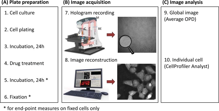
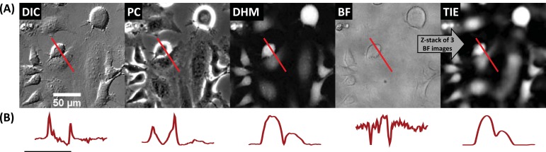
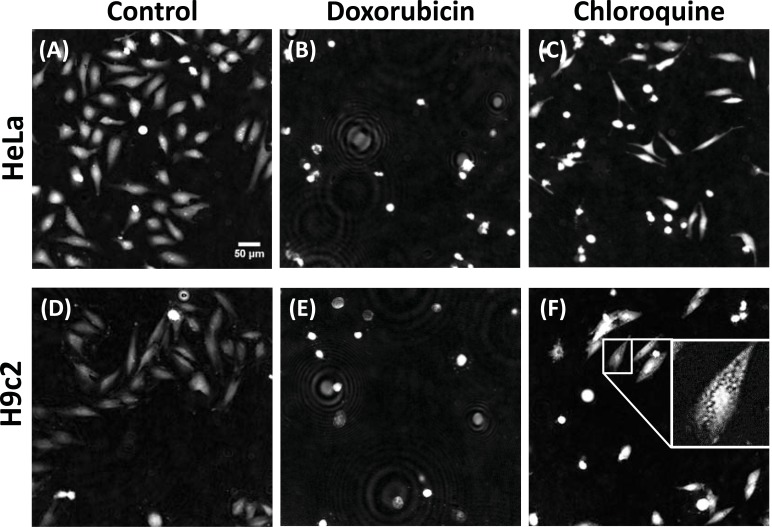
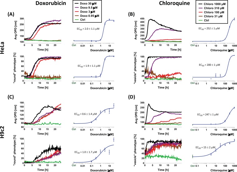
Digital Holographic Microscopy (DHM) is a label-free imaging technique allowing visualization of transparent cells with classical imaging cell culture plates. The quantitative DHM phase contrast image provided is related both to the intracellular refractive index and to cell thickness. DHM is able to distinguish cellular morphological changes on two representative cell lines (HeLa and H9c2) when treated with doxorubicin and chloroquine, two cytotoxic compounds yielding distinct phenotypes. We analyzed parameters linked to cell morphology and to the intracellular content in endpoint measurements and further investigated them with timelapse recording. The results obtained by DHM were compared with other optical label-free microscopy techniques, namely Phase Contrast, Differential Interference Contrast and Transport of Intensity Equation (reconstructed from three bright-field images). For comparative purposes, images were acquired in a common 96-well plate format on the different motorized microscopes. In contrast to the other microscopies assayed, images generated with DHM can be easily quantified using a simple automatized on-the-fly analysis method for discriminating the different phenotypes generated in each cell line. The DHM technology is suitable for the development of robust and unbiased image-based assays.

摘要

数字全息显微镜(DHM)是一种无标记成像技术,可通过传统成像细胞培养板实现透明细胞的可视化。所提供的定量DHM相衬图像与细胞内折射率和细胞厚度均相关。当用阿霉素和氯喹这两种产生不同表型的细胞毒性化合物处理时,DHM能够区分两种代表性细胞系(HeLa和H9c2)的细胞形态变化。我们在终点测量中分析了与细胞形态和细胞内含量相关的参数,并通过延时记录进一步研究了这些参数。将DHM获得的结果与其他无标记光学显微镜技术进行了比较,即相衬显微镜、微分干涉相衬显微镜和强度传输方程(由三张明场图像重建)。为了进行比较,在不同的电动显微镜上以常见的96孔板格式采集图像。与所检测的其他显微镜不同,使用DHM生成的图像可以通过一种简单的自动实时分析方法轻松定量,以区分每个细胞系中产生的不同表型。DHM技术适用于开发强大且无偏差的基于图像的检测方法。

https://cdn.ncbi.nlm.nih.gov/pmc/blobs/ca3a/3894694/2ca152511f9a/CCHTS-17-80_F4.jpg
https://cdn.ncbi.nlm.nih.gov/pmc/blobs/ca3a/3894694/c159d4de940a/CCHTS-17-80_F1.jpg
https://cdn.ncbi.nlm.nih.gov/pmc/blobs/ca3a/3894694/1507c023fd97/CCHTS-17-80_F2.jpg
https://cdn.ncbi.nlm.nih.gov/pmc/blobs/ca3a/3894694/5355fda23252/CCHTS-17-80_F3.jpg
https://cdn.ncbi.nlm.nih.gov/pmc/blobs/ca3a/3894694/2ca152511f9a/CCHTS-17-80_F4.jpg

相似文献

1
Digital holographic microscopy: a quantitative label-free microscopy technique for phenotypic screening.数字全息显微镜:一种用于表型筛选的无标记定量显微镜技术。
Comb Chem High Throughput Screen. 2014 Jan;17(1):80-8. doi: 10.2174/13862073113166660062.
2
A practical criterion for focusing of unstained cell samples using a digital holographic microscope.使用数字全息显微镜聚焦未染色细胞样品的实用标准。
J Microsc. 2020 Aug;279(2):114-122. doi: 10.1111/jmi.12924. Epub 2020 Jun 5.
3
Digital holographic microscopy: a noninvasive contrast imaging technique allowing quantitative visualization of living cells with subwavelength axial accuracy.数字全息显微镜:一种无创对比成像技术,可实现对活细胞的定量可视化,轴向精度达到亚波长级别。
Opt Lett. 2005 Mar 1;30(5):468-70. doi: 10.1364/ol.30.000468.
4
Quantitative phase imaging for cell culture quality control.用于细胞培养质量控制的定量相成像
Cytometry A. 2017 May;91(5):470-481. doi: 10.1002/cyto.a.23082. Epub 2017 Mar 6.
5
Movies of cellular and sub-cellular motion by digital holographic microscopy.通过数字全息显微镜观察细胞和亚细胞运动的影片。
Biomed Eng Online. 2006 Mar 23;5:21. doi: 10.1186/1475-925X-5-21.
6
Label-free quantitative cell division monitoring of endothelial cells by digital holographic microscopy.利用数字全息显微镜对内皮细胞进行无标记定量细胞分裂监测。
J Biomed Opt. 2010 May-Jun;15(3):036009. doi: 10.1117/1.3431712.
7
High space-bandwidth in quantitative phase imaging using partially spatially coherent digital holographic microscopy and a deep neural network.利用部分空间相干数字全息显微镜和深度神经网络实现高空间带宽的定量相位成像。
Opt Express. 2020 Nov 23;28(24):36229-36244. doi: 10.1364/OE.402666.
8
Label-Free Digital Holographic Microscopy for In Vitro Cytotoxic Effect Quantification of Organic Nanoparticles.无标记数字全息显微镜用于体外有机纳米颗粒细胞毒性效应定量分析。
Cells. 2022 Feb 12;11(4):644. doi: 10.3390/cells11040644.
9
Non-invasive, label-free cell counting and quantitative analysis of adherent cells using digital holography.使用数字全息术对贴壁细胞进行无创、无标记的细胞计数和定量分析。
J Microsc. 2008 Nov;232(2):240-7. doi: 10.1111/j.1365-2818.2008.02095.x.
10
Sample and substrate preparation for exploring living neurons in culture with quantitative-phase imaging.用定量相成像技术探索培养活神经元的样品和基质制备。
Methods. 2018 Mar 1;136:90-107. doi: 10.1016/j.ymeth.2018.02.001. Epub 2018 Feb 10.

引用本文的文献

1
Dissecting Morphological and Functional Dynamics of Non-Tumorigenic and Triple-Negative Breast Cancer Cell Lines Using PCA and t-SNE Analysis.使用主成分分析(PCA)和t-分布随机邻域嵌入(t-SNE)分析剖析非致瘤性和三阴性乳腺癌细胞系的形态学和功能动力学
Cancer Rep (Hoboken). 2025 Jul;8(7):e70257. doi: 10.1002/cnr2.70257.
2
Advances in Microscopy and Its Applications with Special Reference to Fluorescence Microscope: An Overview.显微镜技术进展及其应用——特别涉及荧光显微镜:综述
Adv Exp Med Biol. 2023;1406:3-17. doi: 10.1007/978-3-031-26462-7_1.
3
Label-free viability assay using in-line holographic video microscopy.

本文引用的文献

1
A high content assay to assess cellular fitness.一种用于评估细胞健康状况的高内涵分析方法。
Comb Chem High Throughput Screen. 2014 Jan;17(1):12-24. doi: 10.2174/13862073113169990056.
2
Automated segmentation of multiple red blood cells with digital holographic microscopy.利用数字全息显微镜对多个红细胞进行自动分割。
J Biomed Opt. 2013 Feb;18(2):26006. doi: 10.1117/1.JBO.18.2.026006.
3
Label-free cytotoxicity screening assay by digital holographic microscopy.通过数字全息显微镜进行的无标记细胞毒性筛选测定。
无标记细胞活力检测的实时全息视频显微镜法。
Sci Rep. 2022 Jul 26;12(1):12746. doi: 10.1038/s41598-022-17098-y.
4
Nanospermidine in Combination with Nanofenretinide Induces Cell Death in Neuroblastoma Cell Lines.纳米亚精胺与纳米视黄醛联合诱导神经母细胞瘤细胞系细胞死亡。
Pharmaceutics. 2022 Jun 7;14(6):1215. doi: 10.3390/pharmaceutics14061215.
5
Characterization and identification of cell death dynamics by quantitative phase imaging.通过定量相位成像对细胞死亡动力学进行特征化和鉴定。
J Biomed Opt. 2022 Apr;27(4). doi: 10.1117/1.JBO.27.4.046502.
6
Image- and Fluorescence-Based Test Shows Oxidant-Dependent Damages in Red Blood Cells and Enables Screening of Potential Protective Molecules.基于图像和荧光的检测方法可显示红细胞中的氧化性损伤,并可筛选潜在的保护分子。
Int J Mol Sci. 2021 Apr 20;22(8):4293. doi: 10.3390/ijms22084293.
7
High-throughput label-free cell detection and counting from diffraction patterns with deep fully convolutional neural networks.利用深度全卷积神经网络从衍射模式中进行高通量无标记细胞检测和计数。
J Biomed Opt. 2021 Mar;26(3). doi: 10.1117/1.JBO.26.3.036001.
8
Nonmechanical parfocal and autofocus features based on wave propagation distribution in lensfree holographic microscopy.基于无透镜全息显微镜中波传播分布的非机械共焦和自动对焦功能。
Sci Rep. 2021 Feb 5;11(1):3213. doi: 10.1038/s41598-021-81098-7.
9
Root Extracts of Two Cultivars of Species: Lipid Composition and Biological Effects on Different Cell Lines: Preliminary Results.两种 品种根提取物:脂质成分和对不同细胞系的生物学效应:初步结果。
Molecules. 2021 Jan 27;26(3):655. doi: 10.3390/molecules26030655.
10
Acoustofluidic phase microscopy in a tilted segmentation-free configuration.倾斜无分割配置下的声流相显微镜技术。
Biomicrofluidics. 2021 Jan 5;15(1):014102. doi: 10.1063/5.0036585. eCollection 2021 Jan.
Assay Drug Dev Technol. 2013 Mar;11(2):101-7. doi: 10.1089/adt.2012.476. Epub 2012 Oct 12.
4
Understanding the phase contrast optics to restore artifact-free microscopy images for segmentation.理解相衬光学原理,以恢复无伪影的显微镜图像进行分割。
Med Image Anal. 2012 Jul;16(5):1047-62. doi: 10.1016/j.media.2011.12.006. Epub 2012 Feb 3.
5
Early cell death detection with digital holographic microscopy.利用数字全息显微镜检测早期细胞死亡。
PLoS One. 2012;7(1):e30912. doi: 10.1371/journal.pone.0030912. Epub 2012 Jan 31.
6
Determination of transmembrane water fluxes in neurons elicited by glutamate ionotropic receptors and by the cotransporters KCC2 and NKCC1: a digital holographic microscopy study.谷氨酸离子型受体和共转运体 KCC2 和 NKCC1 诱发神经元跨膜水通量的测定:数字全息显微镜研究。
J Neurosci. 2011 Aug 17;31(33):11846-54. doi: 10.1523/JNEUROSCI.0286-11.2011.
7
Quantitative phase restoration by direct inversion using the optical transfer function.使用光学传递函数直接反演进行定量相位恢复。
Opt Lett. 2011 Jul 15;36(14):2671-3. doi: 10.1364/OL.36.002671.
8
Restoring DIC microscopy images from multiple shear directions.从多个剪切方向恢复微分干涉差显微镜图像。
Inf Process Med Imaging. 2011;22:384-97. doi: 10.1007/978-3-642-22092-0_32.
9
A high content screening assay for identifying lysosomotropic compounds.一种用于鉴定溶酶体靶向化合物的高内涵筛选测定法。
Toxicol In Vitro. 2011 Apr;25(3):715-23. doi: 10.1016/j.tiv.2010.12.010. Epub 2010 Dec 22.
10
Differential cytotoxic actions of Shiga toxin 1 and Shiga toxin 2 on microvascular and macrovascular endothelial cells.志贺毒素 1 和志贺毒素 2 对微血管和大血管内皮细胞的细胞毒性作用差异。
Thromb Haemost. 2011 Mar;105(3):515-28. doi: 10.1160/TH10-02-0140. Epub 2010 Dec 6.