Matalia Jyoti, Anegondi Neha Sutheekshna, Veeboy Leio, Roy Abhijit Sinha
Department of Pediatric Ophthalmology, Narayana Nethralaya, Bangalore, India.
School of Biosciences and Technology, VIT University, Vellore, India.
Indian J Ophthalmol. 2018 Jan;66(1):77-82. doi: 10.4103/ijo.IJO_652_17.
To evaluate the association between retinal and choroidal thickness and volume along with choroidal vessel volume in children using optical coherence tomography (OCT) images.
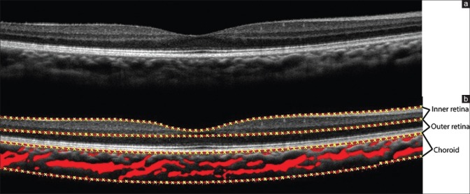
113 normal eyes of children ranging from 5-17 years of age were imaged with a clinical OCT scanner (Optovue Inc., Fremont, USA). The retina, choroid and choroidal vessels were automatically segmented with algorithms. Parameters evaluated were thickness and volume. Location specific analyses of thickness were also performed at a distance of 2.5 mm from foveal center. Multivariate analyses of variance were used to analyze the effect of age and myopia. Manual segmentation of the fovea and subfoveal choroid thickness was also performed to compare with the algorithm segmentation.
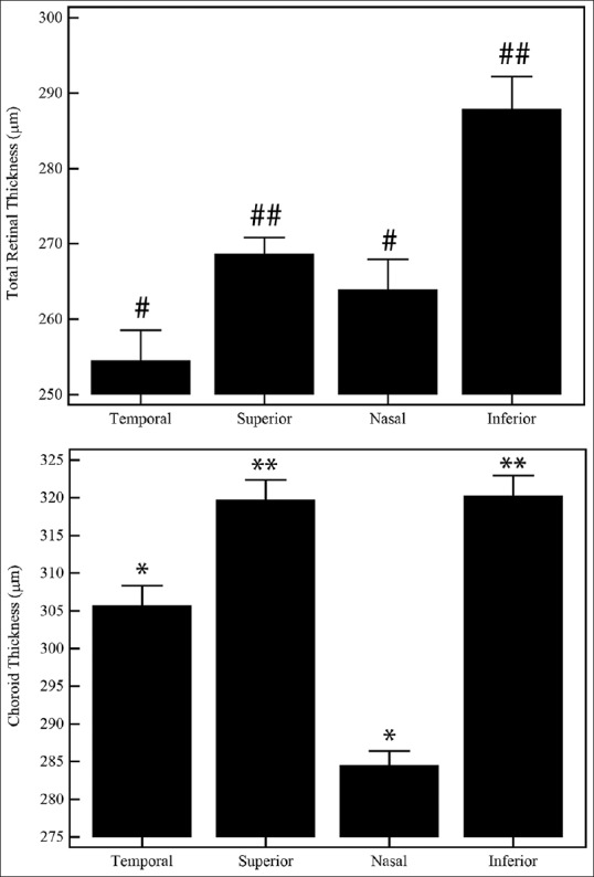
There was excellent agreement between manual and automatic segmentation (intra-class correlation of 0.95). Within the same eye, total retinal and choroid thickness of nasal and temporal location were significantly lower than the superior and inferior thickness (P < 0.0001). With age (P = 0.026) and myopia (P < 0.001), foveal thickness increased. Choroid volume, vessel volume and temporal choroid thickness increased with increasing myopia (P < 0.05). There was significant positive correlation between choroid volume and retinal volume (r = 0.62, P < 0.0001), choroid volume and vessel volume (r = 0.48, P < 0.0001), and with foveal thickness (r = 0.31, P = 0.009). Choroid vessel volume also showed significant positive correlations with the other metrics (P < 0.05).
Retinal and choroidal structural features were quantified simultaneously from OCT images. Magnitude of myopia had a greater effect on retino-choroid features than age in children.
利用光学相干断层扫描(OCT)图像评估儿童视网膜和脉络膜厚度、体积以及脉络膜血管体积之间的关联。
使用临床OCT扫描仪(美国弗里蒙特市Optovue公司)对113只年龄在5至17岁的儿童正常眼睛进行成像。通过算法自动分割视网膜、脉络膜和脉络膜血管。评估的参数为厚度和体积。还在距黄斑中心2.5毫米处进行了厚度的特定位置分析。采用多变量方差分析来分析年龄和近视的影响。还对黄斑和黄斑下脉络膜厚度进行手动分割,以与算法分割结果进行比较。
手动分割与自动分割之间具有极好的一致性(组内相关系数为0.95)。在同一只眼睛内,鼻侧和颞侧位置的视网膜和脉络膜总厚度显著低于上方和下方的厚度(P < 0.0001)。随着年龄增长(P = 0.026)和近视程度增加(P < 0.001),黄斑厚度增加。脉络膜体积、血管体积和颞侧脉络膜厚度随着近视程度增加而增加(P < 0.05)。脉络膜体积与视网膜体积之间存在显著正相关(r = 0.62,P < 0.0001),脉络膜体积与血管体积之间存在显著正相关(r = 0.48,P < 0.0001),与黄斑厚度之间也存在显著正相关(r = 0.31,P = 0.009)。脉络膜血管体积与其他指标也显示出显著正相关(P < 0.05)。
从OCT图像中同时量化了视网膜和脉络膜的结构特征。在儿童中,近视程度对视网膜 - 脉络膜特征的影响大于年龄。