Pshenay-Severin Ekaterina, Bae Hyeonsoo, Reichwald Karl, Matz Gregor, Bierlich Jörg, Kobelke Jens, Lorenz Adrian, Schwuchow Anka, Meyer-Zedler Tobias, Schmitt Michael, Messerschmidt Bernhard, Popp Juergen
GRINTECH GmbH, Schillerstr. 1, 07745, Jena, Germany.
Leibniz Institute of Photonic Technology, Member of Leibniz Health Technologies, Albert-Einstein-Str. 9, 07745, Jena, Germany.
Light Sci Appl. 2021 Oct 5;10(1):207. doi: 10.1038/s41377-021-00648-w.
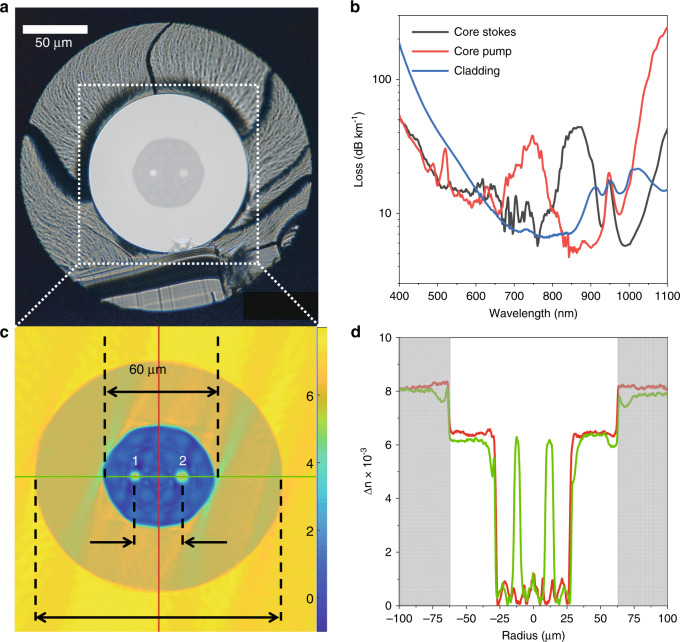
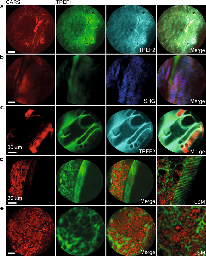
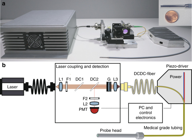
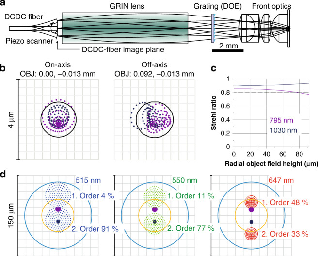
Multimodal non-linear microscopy combining coherent anti-Stokes Raman scattering, second harmonic generation, and two-photon excited fluorescence has proved to be a versatile and powerful tool enabling the label-free investigation of tissue structure, molecular composition, and correlation with function and disease status. For a routine medical application, the implementation of this approach into an in vivo imaging endoscope is required. However, this is a difficult task due to the requirements of a multicolour ultrashort laser delivery from a compact and robust laser source through a fiber with low losses and temporal synchronization, the efficient signal collection in epi-direction, the need for small-diameter but highly corrected endomicroobjectives of high numerical aperture and compact scanners. Here, we introduce an ultra-compact fiber-scanning endoscope platform for multimodal non-linear endomicroscopy in combination with a compact four-wave mixing based fiber laser. The heart of this fiber-scanning endoscope is an in-house custom-designed, single mode, double clad, double core pure silica fiber in combination with a 2.4 mm diameter NIR-dual-waveband corrected endomicroscopic objective of 0.55 numerical aperture and 180 µm field of view for non-linear imaging, allowing a background free, low-loss, high peak power laser delivery, and an efficient signal collection in backward direction. A linear diffractive optical grating overlays pump and Stokes laser foci across the full field of view, such that diffraction-limited performance is demonstrated for tissue imaging at one frame per second with sub-micron spatial resolution and at a high transmission of 65% from the laser to the specimen using a distal resonant fiber scanner.
结合相干反斯托克斯拉曼散射、二次谐波产生和双光子激发荧光的多模态非线性显微镜已被证明是一种通用且强大的工具,能够对组织结构、分子组成以及与功能和疾病状态的相关性进行无标记研究。对于常规医学应用,需要将这种方法应用于体内成像内窥镜。然而,这是一项艰巨的任务,因为需要从紧凑且坚固的激光源通过具有低损耗和时间同步的光纤进行多色超短激光传输,在落射方向上高效收集信号,需要小直径但高度校正的高数值孔径内窥微物镜和紧凑的扫描仪。在此,我们介绍一种超紧凑的光纤扫描内窥镜平台,用于多模态非线性内窥显微镜检查,并结合基于紧凑四波混频的光纤激光器。这种光纤扫描内窥镜的核心是内部定制设计的单模、双包层、双芯纯石英光纤,与一个直径为2.4毫米的近红外双波段校正内窥微物镜相结合,该物镜的数值孔径为0.55,视场为180微米,用于非线性成像,可实现无背景、低损耗、高峰值功率的激光传输,并在向后方向上高效收集信号。一个线性衍射光栅在整个视场上覆盖泵浦和斯托克斯激光焦点,从而使用远端共振光纤扫描仪以每秒一帧的速度对组织成像,展示出具有亚微米空间分辨率且从激光到样本的传输率高达65%的衍射极限性能。