Department of Materials Science and Engineering, Massachusetts Institute of Technology, Cambridge, MA, 02139, USA.
The David H. Koch Institute for Integrative Cancer Research, Massachusetts Institute of Technology, Cambridge, MA, 02139, USA.
Sci Rep. 2019 Mar 7;9(1):3873. doi: 10.1038/s41598-019-39502-w.
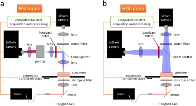
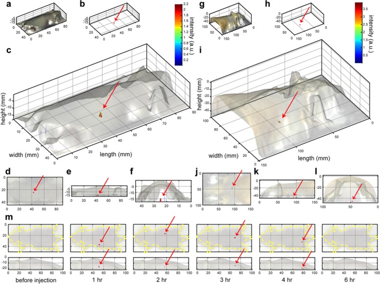
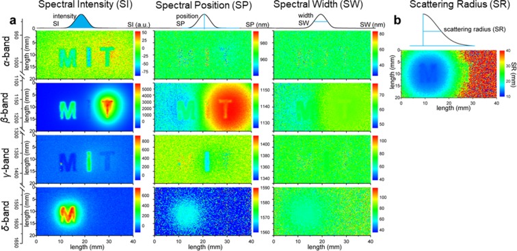
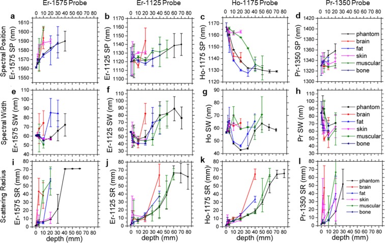
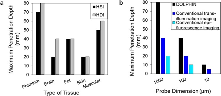
Detection of biological features at the cellular level with sufficient sensitivity in complex tissue remains a major challenge. To appreciate this challenge, this would require finding tens to hundreds of cells (a 0.1 mm tumor has ~125 cells), out of ~37 trillion cells in the human body. Near-infrared optical imaging holds promise for high-resolution, deep-tissue imaging, but is limited by autofluorescence and scattering. To date, the maximum reported depth using second-window near-infrared (NIR-II: 1000-1700 nm) fluorophores is 3.2 cm through tissue. Here, we design an NIR-II imaging system, "Detection of Optically Luminescent Probes using Hyperspectral and diffuse Imaging in Near-infrared" (DOLPHIN), that resolves these challenges. DOLPHIN achieves the following: (i) resolution of probes through up to 8 cm of tissue phantom; (ii) identification of spectral and scattering signatures of tissues without a priori knowledge of background or autofluorescence; and (iii) 3D reconstruction of live whole animals. Notably, we demonstrate noninvasive real-time tracking of a 0.1 mm-sized fluorophore through the gastrointestinal tract of a living mouse, which is beyond the detection limit of current imaging modalities.
在复杂组织中以足够的灵敏度检测细胞水平的生物特征仍然是一个主要挑战。为了了解这一挑战,这需要在人体中找到数十到数百个细胞(一个 0.1 毫米大小的肿瘤有大约 125 个细胞)。近红外光学成像是高分辨率、深层组织成像的有希望的方法,但受到自发荧光和散射的限制。迄今为止,使用第二近红外窗口(NIR-II:1000-1700nm)荧光团报告的最大深度为通过组织的 3.2 厘米。在这里,我们设计了一种近红外成像系统“Detection of Optically Luminescent Probes using Hyperspectral and diffuse Imaging in Near-infrared”(DOLPHIN),可解决这些挑战。DOLPHIN 实现了以下目标:(i)通过高达 8 厘米的组织模型分辨探针;(ii)在没有背景或自发荧光先验知识的情况下识别组织的光谱和散射特征;(iii)对活体动物进行 3D 重建。值得注意的是,我们展示了通过活体小鼠的胃肠道无创实时追踪 0.1 毫米大小的荧光团,这超出了当前成像方式的检测极限。