• 文献检索
  • 文档翻译
  • 深度研究
  • 学术资讯
  • Suppr Zotero 插件Zotero 插件
  • 邀请有礼
  • 套餐&价格
  • 历史记录
应用&插件
Suppr Zotero 插件Zotero 插件浏览器插件Mac 客户端Windows 客户端微信小程序
定价
高级版会员购买积分包购买API积分包
服务
文献检索文档翻译深度研究API 文档MCP 服务
关于我们
关于 Suppr公司介绍联系我们用户协议隐私条款
关注我们

Suppr 超能文献

核心技术专利:CN118964589B侵权必究
粤ICP备2023148730 号-1Suppr @ 2026

文献检索

告别复杂PubMed语法,用中文像聊天一样搜索,搜遍4000万医学文献。AI智能推荐,让科研检索更轻松。

立即免费搜索

文件翻译

保留排版,准确专业,支持PDF/Word/PPT等文件格式,支持 12+语言互译。

免费翻译文档

深度研究

AI帮你快速写综述,25分钟生成高质量综述,智能提取关键信息,辅助科研写作。

立即免费体验

使用3D U-Net集成进行脑肿瘤分割以及使用放射组学特征进行总生存预测

Brain Tumor Segmentation Using an Ensemble of 3D U-Nets and Overall Survival Prediction Using Radiomic Features.

作者信息

Feng Xue, Tustison Nicholas J, Patel Sohil H, Meyer Craig H

机构信息

Department of Biomedical Engineering, University of Virginia, Charlottesville, VA, United States.

Department of Radiology and Medical Imaging, University of Virginia, Charlottesville, VA, United States.

出版信息

Front Comput Neurosci. 2020 Apr 8;14:25. doi: 10.3389/fncom.2020.00025. eCollection 2020.

DOI:10.3389/fncom.2020.00025
PMID:32322196
原文链接:https://pmc.ncbi.nlm.nih.gov/articles/PMC7158872/
Abstract

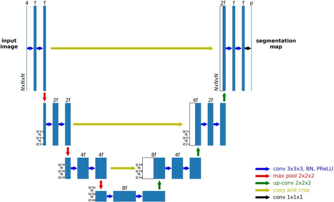
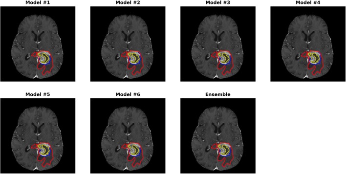
Accurate segmentation of different sub-regions of gliomas such as peritumoral edema, necrotic core, enhancing, and non-enhancing tumor core from multimodal MRI scans has important clinical relevance in diagnosis, prognosis and treatment of brain tumors. However, due to the highly heterogeneous appearance and shape of these tumors, segmentation of the sub-regions is challenging. Recent developments using deep learning models has proved its effectiveness in various semantic and medical image segmentation tasks, many of which are based on the U-Net network structure with symmetric encoding and decoding paths for end-to-end segmentation due to its high efficiency and good performance. In brain tumor segmentation, the 3D nature of multimodal MRI poses challenges such as memory and computation limitations and class imbalance when directly adopting the U-Net structure. In this study we aim to develop a deep learning model using a 3D U-Net with adaptations in the training and testing strategies, network structures, and model parameters for brain tumor segmentation. Furthermore, instead of picking one best model, an ensemble of multiple models trained with different hyper-parameters are used to reduce random errors from each model and yield improved performance. Preliminary results demonstrate the effectiveness of this method and achieved the 9th place in the very competitive 2018 Multimodal Brain Tumor Segmentation (BraTS) challenge. In addition, to emphasize the clinical value of the developed segmentation method, a linear model based on the radiomics features extracted from segmentation and other clinical features are developed to predict patient overall survival. Evaluation of these innovations shows high prediction accuracy in both low-grade glioma and glioblastoma patients, which achieved the 1st place in the 2018 BraTS challenge.

摘要

从多模态磁共振成像(MRI)扫描中准确分割胶质瘤的不同子区域,如瘤周水肿、坏死核心、强化和非强化肿瘤核心,在脑肿瘤的诊断、预后和治疗中具有重要的临床意义。然而,由于这些肿瘤的外观和形状高度异质性,子区域的分割具有挑战性。最近使用深度学习模型的进展已证明其在各种语义和医学图像分割任务中的有效性,其中许多基于具有对称编码和解码路径的U-Net网络结构进行端到端分割,因其效率高和性能好。在脑肿瘤分割中,多模态MRI的三维性质带来了诸如内存和计算限制以及直接采用U-Net结构时的类别不平衡等挑战。在本研究中,我们旨在开发一种深度学习模型,该模型使用3D U-Net,并在训练和测试策略、网络结构和模型参数方面进行调整以用于脑肿瘤分割。此外,不是选择一个最佳模型,而是使用由不同超参数训练的多个模型的集成来减少每个模型的随机误差并提高性能。初步结果证明了该方法的有效性,并在竞争激烈的2018年多模态脑肿瘤分割(BraTS)挑战赛中获得了第9名。此外,为了强调所开发分割方法的临床价值,基于从分割中提取的放射组学特征和其他临床特征开发了一种线性模型来预测患者的总生存期。对这些创新的评估显示,在低级别胶质瘤和胶质母细胞瘤患者中均具有较高的预测准确性,这在2018年BraTS挑战赛中获得了第1名。

https://cdn.ncbi.nlm.nih.gov/pmc/blobs/f959/7158872/b21ebd2c5419/fncom-14-00025-g0007.jpg
https://cdn.ncbi.nlm.nih.gov/pmc/blobs/f959/7158872/e30d70d38102/fncom-14-00025-g0001.jpg
https://cdn.ncbi.nlm.nih.gov/pmc/blobs/f959/7158872/516181a9f441/fncom-14-00025-g0002.jpg
https://cdn.ncbi.nlm.nih.gov/pmc/blobs/f959/7158872/2ea49adabcd2/fncom-14-00025-g0003.jpg
https://cdn.ncbi.nlm.nih.gov/pmc/blobs/f959/7158872/8ea7275785a9/fncom-14-00025-g0004.jpg
https://cdn.ncbi.nlm.nih.gov/pmc/blobs/f959/7158872/500d1173600c/fncom-14-00025-g0005.jpg
https://cdn.ncbi.nlm.nih.gov/pmc/blobs/f959/7158872/b8a5437d331c/fncom-14-00025-g0006.jpg
https://cdn.ncbi.nlm.nih.gov/pmc/blobs/f959/7158872/b21ebd2c5419/fncom-14-00025-g0007.jpg

相似文献

1
Brain Tumor Segmentation Using an Ensemble of 3D U-Nets and Overall Survival Prediction Using Radiomic Features.使用3D U-Net集成进行脑肿瘤分割以及使用放射组学特征进行总生存预测
Front Comput Neurosci. 2020 Apr 8;14:25. doi: 10.3389/fncom.2020.00025. eCollection 2020.
2
Brain Tumor Segmentation and Survival Prediction Using Multimodal MRI Scans With Deep Learning.使用深度学习的多模态磁共振成像扫描进行脑肿瘤分割与生存预测
Front Neurosci. 2019 Aug 16;13:810. doi: 10.3389/fnins.2019.00810. eCollection 2019.
3
A Novel Approach for Fully Automatic Intra-Tumor Segmentation With 3D U-Net Architecture for Gliomas.一种采用3D U-Net架构对胶质瘤进行全自动肿瘤内分割的新方法。
Front Comput Neurosci. 2020 Feb 18;14:10. doi: 10.3389/fncom.2020.00010. eCollection 2020.
4
A radiomics-incorporated deep ensemble learning model for multi-parametric MRI-based glioma segmentation.基于放射组学的深度集成学习模型在多参数 MRI 脑胶质瘤分割中的应用。
Phys Med Biol. 2023 Sep 13;68(18). doi: 10.1088/1361-6560/acf10d.
5
Asymmetric Ensemble of Asymmetric U-Net Models for Brain Tumor Segmentation With Uncertainty Estimation.用于脑肿瘤分割并带有不确定性估计的非对称U-Net模型的非对称集成
Front Neurol. 2021 Sep 30;12:609646. doi: 10.3389/fneur.2021.609646. eCollection 2021.
6
Dilated multi-scale residual attention (DMRA) U-Net: three-dimensional (3D) dilated multi-scale residual attention U-Net for brain tumor segmentation.扩张多尺度残差注意力(DMRA)U-Net:用于脑肿瘤分割的三维(3D)扩张多尺度残差注意力U-Net。
Quant Imaging Med Surg. 2024 Oct 1;14(10):7249-7264. doi: 10.21037/qims-24-779. Epub 2024 Sep 19.
7
Automated glioma grading on conventional MRI images using deep convolutional neural networks.使用深度卷积神经网络对传统MRI图像进行自动脑胶质瘤分级
Med Phys. 2020 Jul;47(7):3044-3053. doi: 10.1002/mp.14168. Epub 2020 May 11.
8
Overall Survival Prediction of Glioma Patients With Multiregional Radiomics.多区域放射组学对胶质瘤患者的总生存预测
Front Neurosci. 2022 Jul 7;16:911065. doi: 10.3389/fnins.2022.911065. eCollection 2022.
9
A novel end-to-end brain tumor segmentation method using improved fully convolutional networks.一种使用改进的全卷积网络的新型端到端脑肿瘤分割方法。
Comput Biol Med. 2019 May;108:150-160. doi: 10.1016/j.compbiomed.2019.03.014. Epub 2019 Mar 18.
10
SGEResU-Net for brain tumor segmentation.基于 SGResU-Net 的脑肿瘤分割。
Math Biosci Eng. 2022 Mar 30;19(6):5576-5590. doi: 10.3934/mbe.2022261.

引用本文的文献

1
OVERALL SURVIVAL PREDICTION OF BRAIN TUMOR PATIENTS WITH MULTIMODAL MRI USING SWIN UNETR.使用Swin UNETR对多模态MRI脑肿瘤患者的总生存预测
Proc IEEE Int Symp Biomed Imaging. 2025 Apr;2025. doi: 10.1109/isbi60581.2025.10981128. Epub 2025 May 12.
2
3D-NASE: A Novel 3D CT Nasal Attention-Based Segmentation Ensemble.3D-NASE:一种基于3D CT鼻腔注意力的新型分割集成方法。
J Imaging. 2025 May 7;11(5):148. doi: 10.3390/jimaging11050148.
3
The clinical implications and interpretability of computational medical imaging (radiomics) in brain tumors.

本文引用的文献

1
Fully Automatic Brain Tumor Segmentation using End-To-End Incremental Deep Neural Networks in MRI images.基于端到端增量式深度神经网络的 MRI 图像全自动脑肿瘤分割。
Comput Methods Programs Biomed. 2018 Nov;166:39-49. doi: 10.1016/j.cmpb.2018.09.007. Epub 2018 Sep 21.
2
A Multi-scale U-Net for Semantic Segmentation of Histological Images from Radical Prostatectomies.用于根治性前列腺切除组织学图像语义分割的多尺度U-Net
AMIA Annu Symp Proc. 2018 Apr 16;2017:1140-1148. eCollection 2017.
3
Brain tumor segmentation in multi-spectral MRI using convolutional neural networks (CNN).
计算医学成像(影像组学)在脑肿瘤中的临床意义及可解释性。
Insights Imaging. 2025 Mar 30;16(1):77. doi: 10.1186/s13244-025-01950-6.
4
Context aware machine learning techniques for brain tumor classification and detection - A review.用于脑肿瘤分类与检测的上下文感知机器学习技术——综述
Heliyon. 2025 Jan 13;11(2):e41835. doi: 10.1016/j.heliyon.2025.e41835. eCollection 2025 Jan 30.
5
EXACT-Net: Framework for EHR-Guided Lung Tumor Auto-Segmentation for Non-Small Cell Lung Cancer Radiotherapy.EXACT-Net:用于非小细胞肺癌放疗的电子健康记录引导下肺肿瘤自动分割框架
Cancers (Basel). 2024 Dec 6;16(23):4097. doi: 10.3390/cancers16234097.
6
Machine learning based ultrasomics noninvasive predicting EGFR expression status in hepatocellular carcinoma patients.基于机器学习的超声组学无创预测肝细胞癌患者表皮生长因子受体(EGFR)表达状态
Front Med (Lausanne). 2024 Nov 19;11:1483291. doi: 10.3389/fmed.2024.1483291. eCollection 2024.
7
MRI denoising with a non-blind deep complex-valued convolutional neural network.基于非盲复值卷积神经网络的 MRI 去噪。
NMR Biomed. 2025 Jan;38(1):e5291. doi: 10.1002/nbm.5291. Epub 2024 Nov 11.
8
Leveraging State-of-the-Art AI Algorithms in Personalized Oncology: From Transcriptomics to Treatment.在个性化肿瘤学中利用最先进的人工智能算法:从转录组学到治疗
Diagnostics (Basel). 2024 Sep 29;14(19):2174. doi: 10.3390/diagnostics14192174.
9
AeroPath: An airway segmentation benchmark dataset with challenging pathology and baseline method.AeroPath:一个具有挑战性病理情况和基线方法的气道分割基准数据集。
PLoS One. 2024 Oct 2;19(10):e0311416. doi: 10.1371/journal.pone.0311416. eCollection 2024.
10
Two decades of progress in glioma methylation research: the rise of temozolomide resistance and immunotherapy insights.胶质瘤甲基化研究二十年进展:替莫唑胺耐药性的出现及免疫治疗见解
Front Neurosci. 2024 Sep 2;18:1440756. doi: 10.3389/fnins.2024.1440756. eCollection 2024.
使用卷积神经网络(CNN)进行多光谱磁共振成像中的脑肿瘤分割。
Microsc Res Tech. 2018 Apr;81(4):419-427. doi: 10.1002/jemt.22994. Epub 2018 Jan 22.
4
Advancing The Cancer Genome Atlas glioma MRI collections with expert segmentation labels and radiomic features.利用专家分割标签和放射组学特征推进癌症基因组图谱胶质细胞瘤 MRI 数据集。
Sci Data. 2017 Sep 5;4:170117. doi: 10.1038/sdata.2017.117.
5
Deep Learning for Brain MRI Segmentation: State of the Art and Future Directions.用于脑磁共振成像分割的深度学习:现状与未来方向。
J Digit Imaging. 2017 Aug;30(4):449-459. doi: 10.1007/s10278-017-9983-4.
6
Efficient multi-scale 3D CNN with fully connected CRF for accurate brain lesion segmentation.高效多尺度 3D CNN 结合全连接条件随机场实现精准脑损伤分割。
Med Image Anal. 2017 Feb;36:61-78. doi: 10.1016/j.media.2016.10.004. Epub 2016 Oct 29.
7
Brain tumor segmentation with Deep Neural Networks.基于深度神经网络的脑肿瘤分割。
Med Image Anal. 2017 Jan;35:18-31. doi: 10.1016/j.media.2016.05.004. Epub 2016 May 19.
8
The 2016 World Health Organization Classification of Tumors of the Central Nervous System: a summary.2016 年世界卫生组织中枢神经系统肿瘤分类:概述。
Acta Neuropathol. 2016 Jun;131(6):803-20. doi: 10.1007/s00401-016-1545-1. Epub 2016 May 9.
9
Radiomics: Images Are More than Pictures, They Are Data.放射组学:图像不止是图片,它们是数据。
Radiology. 2016 Feb;278(2):563-77. doi: 10.1148/radiol.2015151169. Epub 2015 Nov 18.
10
The Multimodal Brain Tumor Image Segmentation Benchmark (BRATS).多模态脑肿瘤图像分割基准(BRATS)。
IEEE Trans Med Imaging. 2015 Oct;34(10):1993-2024. doi: 10.1109/TMI.2014.2377694. Epub 2014 Dec 4.