Jusuf Nelva Karmila, Putra Imam Budi, Sari Lovena
Department of Dermatology and Venerology, Faculty of Medicine, Universitas Sumatera Utara, Medan, Indonesia.
Clin Cosmet Investig Dermatol. 2020 Oct 22;13:773-780. doi: 10.2147/CCID.S272334. eCollection 2020.
Bacterial activity and inflammation both influence acne vulgaris (AV) formation. is considered as an actor involved in inflammation of AV. Besides , other microbiomes found in AV may also play a role in the pathogenesis. This research was conducted to overview microbiomes found in non-inflammatory and inflammatory lesions of AV.
An observational descriptive study with cross-sectional approach was designed. Sample collection was performed with 40 subjects with AV. In every patient, both non-inflammatory (closed comedone) and inflammatory (pustule) lesion samples were collected by swab. Afterward, bacterial culture was performed, continued by bacterial identification.
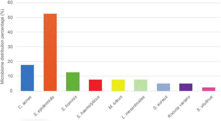
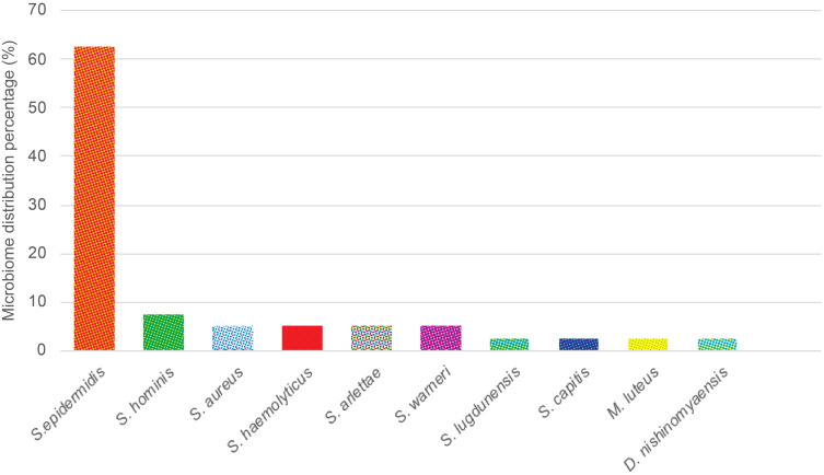
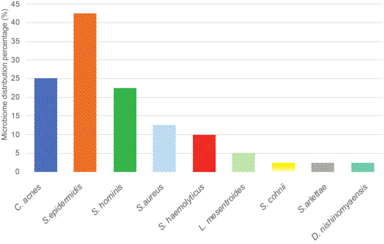
In non-inflammatory lesions, the growth of nine bacterial species was observed from 40 samples. In an anaerobic culture, (17,5%) was identified. In aerobic cultures, different bacterial species were found including (52.5%), (12.5%), (7.5%), (7.5%), (7.5%), (5%), (5%), and (2.5%). In inflammatory lesions, nine bacterial species were found, in which was the anaerobic culture we identified (25.0%). Aerobic cultures have revealed the growth colonies of (42.5%), (22.5%), (12.5%), (10.0%), (5.0%), (2.5%), (2.5%), and (2.5%). Two mixed bacterial growths were observed in non-inflammatory lesions, while four mixed bacterial growths were found in inflammatory lesions.
Differences in bacterial isolates were observed both in non-inflammatory and inflammatory lesions of AV.
细菌活性和炎症均影响寻常痤疮(AV)的形成。 被认为是参与AV炎症的一个因素。此外,在AV中发现的其他微生物群也可能在发病机制中起作用。本研究旨在概述在AV的非炎症性和炎症性病变中发现的微生物群。
设计了一项采用横断面方法的观察性描述性研究。对40例AV患者进行样本采集。在每位患者中,通过拭子采集非炎症性(闭合性粉刺)和炎症性(脓疱)病变样本。随后进行细菌培养,并继续进行细菌鉴定。
在非炎症性病变中,从40个样本中观察到9种细菌的生长。在厌氧培养中,鉴定出 (17.5%)。在需氧培养中,发现了不同的细菌种类,包括 (52.5%)、 (12.5%)、 (7.5%)、 (7.5%)、 (7.5%)、 (5%)、 (5%)和 (2.5%)。在炎症性病变中,发现了9种细菌,其中在厌氧培养中我们鉴定出 (25.0%)。需氧培养显示出 (42.5%)、 (22.5%)、 (12.5%)、 (10.0%)、 (5.0%)、 (2.5%)、 (2.5%)和 (2.5%)的生长菌落。在非炎症性病变中观察到两种混合细菌生长,而在炎症性病变中发现了四种混合细菌生长。
在AV的非炎症性和炎症性病变中均观察到细菌分离株的差异。