Department of Pathology, School of Medicine, College of Medicine, Taipei Medical University, Taipei, Taiwan.
Department of Pathology, Taipei Medical University Hospital, Taipei, Taiwan.
Nat Commun. 2021 Feb 19;12(1):1193. doi: 10.1038/s41467-021-21467-y.
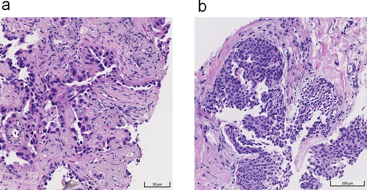
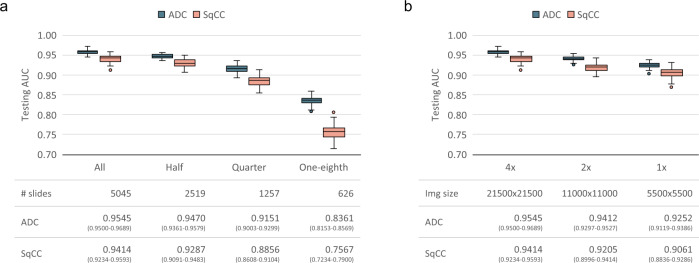
Deep learning for digital pathology is hindered by the extremely high spatial resolution of whole-slide images (WSIs). Most studies have employed patch-based methods, which often require detailed annotation of image patches. This typically involves laborious free-hand contouring on WSIs. To alleviate the burden of such contouring and obtain benefits from scaling up training with numerous WSIs, we develop a method for training neural networks on entire WSIs using only slide-level diagnoses. Our method leverages the unified memory mechanism to overcome the memory constraint of compute accelerators. Experiments conducted on a data set of 9662 lung cancer WSIs reveal that the proposed method achieves areas under the receiver operating characteristic curve of 0.9594 and 0.9414 for adenocarcinoma and squamous cell carcinoma classification on the testing set, respectively. Furthermore, the method demonstrates higher classification performance than multiple-instance learning as well as strong localization results for small lesions through class activation mapping.
深度学习在数字病理学中受到全切片图像(WSI)极高空间分辨率的阻碍。大多数研究都采用基于补丁的方法,而这些方法往往需要对图像补丁进行详细注释。这通常涉及到在 WSI 上进行费力的徒手轮廓绘制。为了减轻这种轮廓绘制的负担,并从使用大量 WSI 进行的培训扩展中获益,我们开发了一种仅使用幻灯片级诊断在整个 WSI 上训练神经网络的方法。我们的方法利用统一的存储机制来克服计算加速器的内存限制。在一个包含 9662 张肺癌 WSI 的数据集上进行的实验表明,所提出的方法在测试集上分别实现了腺癌和鳞状细胞癌分类的受试者工作特征曲线下面积为 0.9594 和 0.9414。此外,该方法通过类激活映射展示了比多实例学习更高的分类性能,以及对小病变的强定位结果。