• 文献检索
  • 文档翻译
  • 深度研究
  • 学术资讯
  • Suppr Zotero 插件Zotero 插件
  • 邀请有礼
  • 套餐&价格
  • 历史记录
应用&插件
Suppr Zotero 插件Zotero 插件浏览器插件Mac 客户端Windows 客户端微信小程序
定价
高级版会员购买积分包购买API积分包
服务
文献检索文档翻译深度研究API 文档MCP 服务
关于我们
关于 Suppr公司介绍联系我们用户协议隐私条款
关注我们

Suppr 超能文献

核心技术专利:CN118964589B侵权必究
粤ICP备2023148730 号-1Suppr @ 2026

文献检索

告别复杂PubMed语法,用中文像聊天一样搜索,搜遍4000万医学文献。AI智能推荐,让科研检索更轻松。

立即免费搜索

文件翻译

保留排版,准确专业,支持PDF/Word/PPT等文件格式,支持 12+语言互译。

免费翻译文档

深度研究

AI帮你快速写综述,25分钟生成高质量综述,智能提取关键信息,辅助科研写作。

立即免费体验

密涅瓦:一种用于多重组织图像的轻量级叙事图像浏览器。

Minerva: a light-weight, narrative image browser for multiplexed tissue images.

作者信息

Hoffer John, Rashid Rumana, Muhlich Jeremy L, Chen Yu-An, Russell Douglas Peter William, Ruokonen Juha, Krueger Robert, Pfister Hanspeter, Santagata Sandro, Sorger Peter K

机构信息

Laboratory of Systems Pharmacology, Harvard Medical School, Boston, MA.

Ludwig Center for Cancer Research at Harvard, Harvard Medical School, Boston, MA.

出版信息

J Open Source Softw. 2020;5(54). doi: 10.21105/joss.02579. Epub 2020 Oct 15.

DOI:10.21105/joss.02579
PMID:33768192
原文链接:https://pmc.ncbi.nlm.nih.gov/articles/PMC7989801/
Abstract

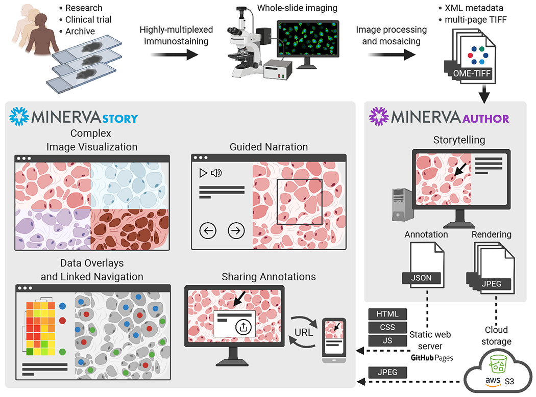
Advances in highly multiplexed tissue imaging are transforming our understanding of human biology by enabling detection and localization of 10-100 proteins at subcellular resolution (Bodenmiller, 2016). Efforts are now underway to create public atlases of multiplexed images of normal and diseased tissues (Rozenblatt-Rosen et al., 2020). Both research and clinical applications of tissue imaging benefit from recording data from complete specimens so that data on cell state and composition can be studied in the context of overall tissue architecture. As a practical matter, specimen size is limited by the dimensions of microscopy slides (2.5 × 7.5 cm or ~2-8 cm of tissue depending on shape). With current microscopy technology, specimens of this size can be imaged at sub-micron resolution across ~60 spectral channels and ~10 cells, resulting in image files of terabyte size. However, the rich detail and multiscale properties of these images pose a substantial computational challenge (Rashid et al., 2020). See Rashid et al. (2020) for an comparison of existing visualization tools targeting these multiplexed tissue images.

摘要

高度多重组织成像技术的进步正在改变我们对人类生物学的理解,它能够在亚细胞分辨率下检测和定位10至100种蛋白质(博登米勒,2016年)。目前正在努力创建正常和患病组织的多重图像公共图谱(罗森布拉特-罗森等人,2020年)。组织成像的研究和临床应用都受益于记录完整标本的数据,以便能够在整体组织结构的背景下研究细胞状态和组成的数据。实际上,标本大小受到显微镜载玻片尺寸的限制(2.5×7.5厘米,或根据形状约为2至8厘米的组织)。使用当前的显微镜技术,这种大小的标本可以在约60个光谱通道和约10个细胞上以亚微米分辨率成像,从而产生数TB大小的图像文件。然而,这些图像丰富的细节和多尺度特性带来了巨大的计算挑战(拉希德等人,2020年)。有关针对这些多重组织图像的现有可视化工具的比较,请参阅拉希德等人(2020年)的文章。

https://cdn.ncbi.nlm.nih.gov/pmc/blobs/b940/7989801/3849bdf1ed16/nihms-1648136-f0002.jpg
https://cdn.ncbi.nlm.nih.gov/pmc/blobs/b940/7989801/c358a2641292/nihms-1648136-f0001.jpg
https://cdn.ncbi.nlm.nih.gov/pmc/blobs/b940/7989801/3849bdf1ed16/nihms-1648136-f0002.jpg
https://cdn.ncbi.nlm.nih.gov/pmc/blobs/b940/7989801/c358a2641292/nihms-1648136-f0001.jpg
https://cdn.ncbi.nlm.nih.gov/pmc/blobs/b940/7989801/3849bdf1ed16/nihms-1648136-f0002.jpg

相似文献

1
Minerva: a light-weight, narrative image browser for multiplexed tissue images.密涅瓦:一种用于多重组织图像的轻量级叙事图像浏览器。
J Open Source Softw. 2020;5(54). doi: 10.21105/joss.02579. Epub 2020 Oct 15.
2
mxnorm: An R Package to Normalize Multiplexed Imaging Data.mxnorm:一个用于标准化多路复用成像数据的R软件包。
J Open Source Softw. 2022;7(71). doi: 10.21105/joss.04180. Epub 2022 Mar 30.
3
In Vivo Observations of Rapid Scattered Light Changes Associated with Neurophysiological Activity与神经生理活动相关的快速散射光变化的体内观察
4
Constructing and optimizing 3D atlases from 2D data with application to the developing mouse brain.从二维数据构建和优化 3D 图谱及其在发育中老鼠大脑中的应用。
Elife. 2021 Feb 11;10:e61408. doi: 10.7554/eLife.61408.
5
Translational Metabolomics of Head Injury: Exploring Dysfunctional Cerebral Metabolism with Ex Vivo NMR Spectroscopy-Based Metabolite Quantification头部损伤的转化代谢组学:基于体外核磁共振波谱的代谢物定量分析探索脑代谢功能障碍
6
Erratum: Eyestalk Ablation to Increase Ovarian Maturation in Mud Crabs.勘误:切除眼柄以增加泥蟹的卵巢成熟度。
J Vis Exp. 2023 May 26(195). doi: 10.3791/6561.
7
Narrative online guides for the interpretation of digital-pathology images and tissue-atlas data.用于解读数字病理学图像和组织图谱数据的叙事式在线指南。
Nat Biomed Eng. 2022 May;6(5):515-526. doi: 10.1038/s41551-021-00789-8. Epub 2021 Nov 8.
8
Highly multiplexed immunofluorescence imaging of human tissues and tumors using t-CyCIF and conventional optical microscopes.使用 t-CyCIF 和传统光学显微镜对人组织和肿瘤进行高多重免疫荧光成像。
Elife. 2018 Jul 11;7:e31657. doi: 10.7554/eLife.31657.
9
Showing their true colors: a practical approach to volume rendering from serial sections.展现真面目:一种从连续切片进行体绘制的实用方法。
BMC Dev Biol. 2010 Apr 21;10:41. doi: 10.1186/1471-213X-10-41.
10
Optical endomicroscopy and the road to real-time, in vivo pathology: present and future.光学内窥和实时体内病理学的道路:现在和未来。
Diagn Pathol. 2012 Aug 13;7:98. doi: 10.1186/1746-1596-7-98.

引用本文的文献

1
Spatial determinants of tumor cell dedifferentiation and plasticity in primary cutaneous melanoma.原发性皮肤黑色素瘤中肿瘤细胞去分化和可塑性的空间决定因素
bioRxiv. 2025 Jun 24:2025.06.21.660851. doi: 10.1101/2025.06.21.660851.
2
Multimodal spatial proteomic profiling in acute myeloid leukemia.急性髓系白血病中的多模态空间蛋白质组学分析
NPJ Precis Oncol. 2025 May 20;9(1):148. doi: 10.1038/s41698-025-00897-7.
3
Sharing data from the Human Tumor Atlas Network through standards, infrastructure and community engagement.通过标准、基础设施和社区参与来分享人类肿瘤图谱网络的数据。

本文引用的文献

1
Narrative online guides for the interpretation of digital-pathology images and tissue-atlas data.用于解读数字病理学图像和组织图谱数据的叙事式在线指南。
Nat Biomed Eng. 2022 May;6(5):515-526. doi: 10.1038/s41551-021-00789-8. Epub 2021 Nov 8.
2
The Human Tumor Atlas Network: Charting Tumor Transitions across Space and Time at Single-Cell Resolution.人类肿瘤图谱网络:以单细胞分辨率绘制肿瘤在空间和时间上的转变图谱。
Cell. 2020 Apr 16;181(2):236-249. doi: 10.1016/j.cell.2020.03.053.
3
Multiplexed Epitope-Based Tissue Imaging for Discovery and Healthcare Applications.
Nat Methods. 2025 Apr;22(4):664-671. doi: 10.1038/s41592-025-02643-0. Epub 2025 Mar 31.
4
Spatial analysis by current multiplexed imaging technologies for the molecular characterisation of cancer tissues.利用当前的多重成像技术进行空间分析,以对癌症组织进行分子特征分析。
Br J Cancer. 2024 Dec;131(11):1737-1747. doi: 10.1038/s41416-024-02882-6. Epub 2024 Oct 22.
5
A perspective on FAIR quality control in multiplexed imaging data processing.多重成像数据处理中FAIR质量控制的观点
Front Bioinform. 2024 Feb 9;4:1336257. doi: 10.3389/fbinf.2024.1336257. eCollection 2024.
6
Multiplexed 3D Analysis of Immune States and Niches in Human Tissue.人体组织中免疫状态和微环境的多重三维分析
bioRxiv. 2024 Mar 28:2023.11.10.566670. doi: 10.1101/2023.11.10.566670.
7
Lymphocyte networks are dynamic cellular communities in the immunoregulatory landscape of lung adenocarcinoma.淋巴细胞网络是肺腺癌免疫调节景观中的动态细胞群落。
Cancer Cell. 2023 May 8;41(5):871-886.e10. doi: 10.1016/j.ccell.2023.03.015. Epub 2023 Apr 13.
8
Interoperable slide microscopy viewer and annotation tool for imaging data science and computational pathology.可互操作的幻灯片显微镜查看器和注释工具,用于成像数据科学和计算病理学。
Nat Commun. 2023 Mar 22;14(1):1572. doi: 10.1038/s41467-023-37224-2.
9
Multiplexed 3D atlas of state transitions and immune interaction in colorectal cancer.多指标 3D 图谱描绘结直肠癌中状态转变和免疫相互作用。
Cell. 2023 Jan 19;186(2):363-381.e19. doi: 10.1016/j.cell.2022.12.028.
10
Spatial omics technologies at multimodal and single cell/subcellular level.空间组学技术在多模态和单细胞/亚细胞水平上的应用。
Genome Biol. 2022 Dec 13;23(1):256. doi: 10.1186/s13059-022-02824-6.
基于多重表位的组织成像在发现和医疗保健中的应用。
Cell Syst. 2016 Apr 27;2(4):225-38. doi: 10.1016/j.cels.2016.03.008.
4
The cBio cancer genomics portal: an open platform for exploring multidimensional cancer genomics data.cBio 癌症基因组学门户:一个用于探索多维癌症基因组学数据的开放平台。
Cancer Discov. 2012 May;2(5):401-4. doi: 10.1158/2159-8290.CD-12-0095.