• 文献检索
  • 文档翻译
  • 深度研究
  • 学术资讯
  • Suppr Zotero 插件Zotero 插件
  • 邀请有礼
  • 套餐&价格
  • 历史记录
应用&插件
Suppr Zotero 插件Zotero 插件浏览器插件Mac 客户端Windows 客户端微信小程序
定价
高级版会员购买积分包购买API积分包
服务
文献检索文档翻译深度研究API 文档MCP 服务
关于我们
关于 Suppr公司介绍联系我们用户协议隐私条款
关注我们

Suppr 超能文献

核心技术专利:CN118964589B侵权必究
粤ICP备2023148730 号-1Suppr @ 2026

文献检索

告别复杂PubMed语法,用中文像聊天一样搜索,搜遍4000万医学文献。AI智能推荐,让科研检索更轻松。

立即免费搜索

文件翻译

保留排版,准确专业,支持PDF/Word/PPT等文件格式,支持 12+语言互译。

免费翻译文档

深度研究

AI帮你快速写综述,25分钟生成高质量综述,智能提取关键信息,辅助科研写作。

立即免费体验

COVID TV-Unet:使用带连通性约束的Unet分割新冠肺炎胸部CT图像

COVID TV-Unet: Segmenting COVID-19 chest CT images using connectivity imposed Unet.

作者信息

Saeedizadeh Narges, Minaee Shervin, Kafieh Rahele, Yazdani Shakib, Sonka Milan

机构信息

Medical Image and Signal Processing Research Center, Isfahan University of Medical Sciences, Iran.

Snap Inc., Seattle, WA, USA.

出版信息

Comput Methods Programs Biomed Update. 2021;1:100007. doi: 10.1016/j.cmpbup.2021.100007. Epub 2021 Apr 20.

DOI:10.1016/j.cmpbup.2021.100007
PMID:34337587
原文链接:https://pmc.ncbi.nlm.nih.gov/articles/PMC8056883/
Abstract

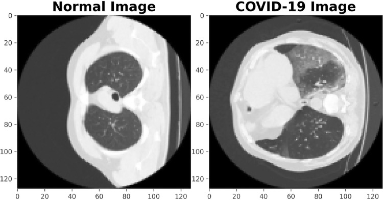
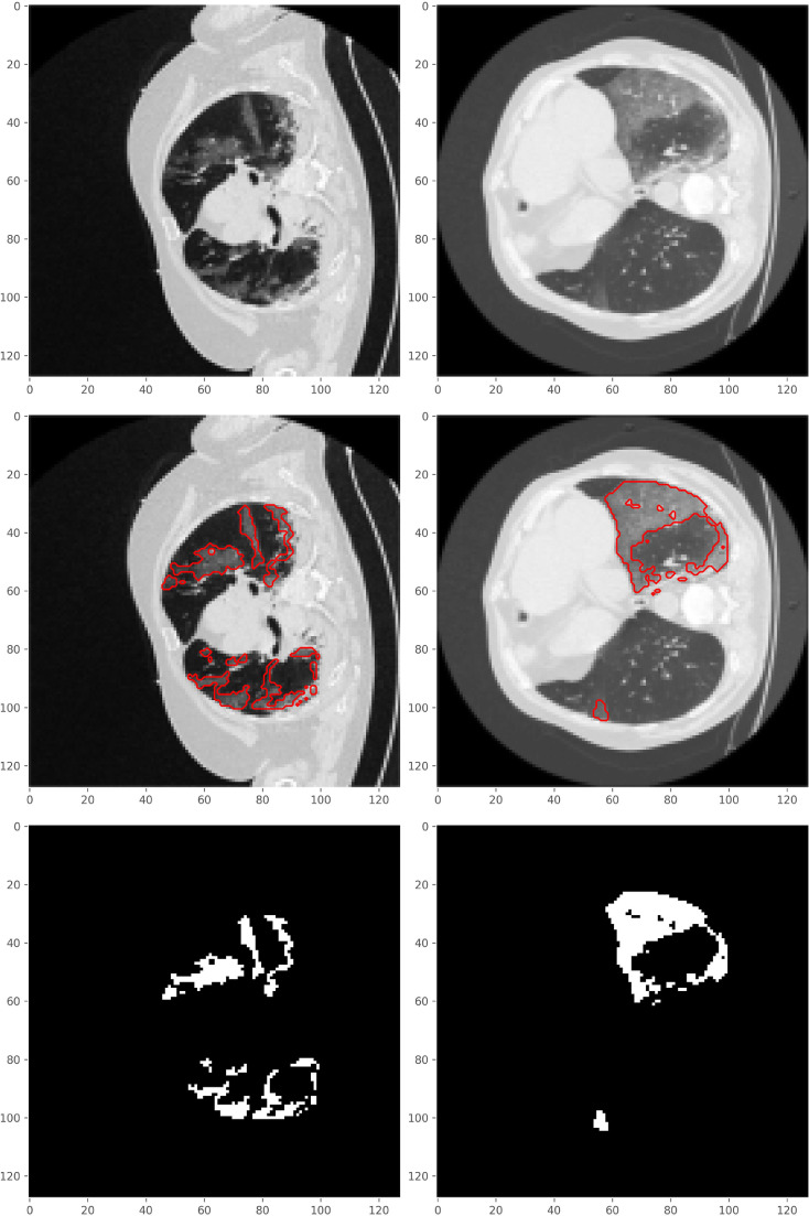
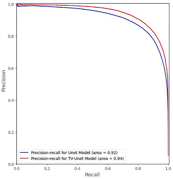
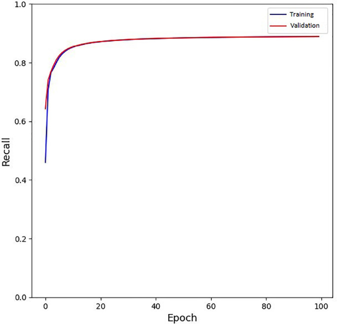
The novel corona-virus disease (COVID-19) pandemic has caused a major outbreak in more than 200 countries around the world, leading to a severe impact on the health and life of many people globally. By October 2020, more than 44 million people were infected, and more than 1,000,000 deaths were reported. Computed Tomography (CT) images can be used as an alternative to the time-consuming RT-PCR test, to detect COVID-19. In this work we propose a segmentation framework to detect chest regions in CT images, which are infected by COVID-19. An architecture similar to a Unet model was employed to detect ground glass regions on a voxel level. As the infected regions tend to form connected components (rather than randomly distributed voxels), a suitable regularization term based on 2D-anisotropic total-variation was developed and added to the loss function. The proposed model is therefore called "TV-Unet". Experimental results obtained on a relatively large-scale CT segmentation dataset of around 900 images, incorporating this new regularization term leads to a 2% gain on overall segmentation performance compared to the Unet trained from scratch. Our experimental analysis, ranging from visual evaluation of the predicted segmentation results to quantitative assessment of segmentation performance (precision, recall, Dice score, and mIoU) demonstrated great ability to identify COVID-19 associated regions of the lungs, achieving a mIoU rate of over 99%, and a Dice score of around 86%.

摘要

新型冠状病毒病(COVID-19)大流行已在全球200多个国家引发大规模疫情,对全球许多人的健康和生活造成了严重影响。截至2020年10月,超过4400万人感染,报告死亡人数超过100万。计算机断层扫描(CT)图像可作为耗时的逆转录聚合酶链反应(RT-PCR)检测的替代方法,用于检测COVID-19。在这项工作中,我们提出了一个分割框架,用于检测CT图像中受COVID-19感染的胸部区域。采用了一种类似于Unet模型的架构,在体素级别上检测磨玻璃区域。由于受感染区域倾向于形成连通分量(而不是随机分布的体素),因此开发了一种基于二维各向异性全变差的合适正则化项,并将其添加到损失函数中。因此,所提出的模型被称为“TV-Unet”。在一个包含约900幅图像的相对大规模CT分割数据集上获得的实验结果表明,与从头开始训练的Unet相比,加入这个新的正则化项可使整体分割性能提高2%。我们的实验分析,从对预测分割结果的视觉评估到分割性能的定量评估(精度、召回率、Dice分数和平均交并比),都显示出该模型具有很强的识别肺部COVID-19相关区域的能力,平均交并比超过99%,Dice分数约为86%。

https://cdn.ncbi.nlm.nih.gov/pmc/blobs/ee8c/8056883/5c6466641a23/gr9_lrg.jpg
https://cdn.ncbi.nlm.nih.gov/pmc/blobs/ee8c/8056883/e352e745f833/gr1_lrg.jpg
https://cdn.ncbi.nlm.nih.gov/pmc/blobs/ee8c/8056883/cfec8a25a2bd/gr2_lrg.jpg
https://cdn.ncbi.nlm.nih.gov/pmc/blobs/ee8c/8056883/5ed3ece65c62/gr3_lrg.jpg
https://cdn.ncbi.nlm.nih.gov/pmc/blobs/ee8c/8056883/0b6615ef7e52/gr4_lrg.jpg
https://cdn.ncbi.nlm.nih.gov/pmc/blobs/ee8c/8056883/bfeef84b4fd1/gr5_lrg.jpg
https://cdn.ncbi.nlm.nih.gov/pmc/blobs/ee8c/8056883/a959869551e7/gr6_lrg.jpg
https://cdn.ncbi.nlm.nih.gov/pmc/blobs/ee8c/8056883/d04f37abe074/gr7_lrg.jpg
https://cdn.ncbi.nlm.nih.gov/pmc/blobs/ee8c/8056883/22a2c190676c/gr8_lrg.jpg
https://cdn.ncbi.nlm.nih.gov/pmc/blobs/ee8c/8056883/5c6466641a23/gr9_lrg.jpg

相似文献

1
COVID TV-Unet: Segmenting COVID-19 chest CT images using connectivity imposed Unet.COVID TV-Unet:使用带连通性约束的Unet分割新冠肺炎胸部CT图像
Comput Methods Programs Biomed Update. 2021;1:100007. doi: 10.1016/j.cmpbup.2021.100007. Epub 2021 Apr 20.
2
COVID TV-UNet: Segmenting COVID-19 Chest CT Images Using Connectivity Imposed U-Net.COVID TV-UNet:使用施加连通性的U-Net分割新冠肺炎胸部CT图像
ArXiv. 2020 Aug 6:arXiv:2007.12303v3.
3
Automated deep learning-based segmentation of COVID-19 lesions from chest computed tomography images.基于深度学习的胸部计算机断层扫描图像中新冠病毒肺炎病变的自动分割
Pol J Radiol. 2022 Aug 26;87:e478-e486. doi: 10.5114/pjr.2022.119027. eCollection 2022.
4
Dual attention fusion UNet for COVID-19 lesion segmentation from CT images.基于双注意力融合 U-Net 的 CT 图像新冠肺炎病灶分割。
J Xray Sci Technol. 2023;31(4):713-729. doi: 10.3233/XST-230001.
5
Automatic COVID-19 Lung Infection Segmentation through Modified Unet Model.通过改进的 U-Net 模型进行自动 COVID-19 肺部感染分割。
J Healthc Eng. 2022 Apr 11;2022:6566982. doi: 10.1155/2022/6566982. eCollection 2022.
6
Enhancing COVID-19 CT Image Segmentation: A Comparative Study of Attention and Recurrence in UNet Models.增强新冠病毒CT图像分割:UNet模型中注意力机制与循环机制的比较研究
J Imaging. 2023 Dec 18;9(12):283. doi: 10.3390/jimaging9120283.
7
Decoders configurations based on Unet family and feature pyramid network for COVID-19 segmentation on CT images.基于Unet家族和特征金字塔网络的解码器配置,用于CT图像上的新冠肺炎分割。
PeerJ Comput Sci. 2021 Sep 17;7:e719. doi: 10.7717/peerj-cs.719. eCollection 2021.
8
CARes-UNet: Content-aware residual UNet for lesion segmentation of COVID-19 from chest CT images.CARes-UNet:用于胸部 CT 图像中 COVID-19 病变分割的基于内容感知残差 UNet 模型。
Med Phys. 2021 Nov;48(11):7127-7140. doi: 10.1002/mp.15231. Epub 2021 Sep 25.
9
MID-UNet: Multi-input directional UNet for COVID-19 lung infection segmentation from CT images.MID-UNet:用于从CT图像中分割新冠病毒肺部感染区域的多输入定向UNet
Signal Process Image Commun. 2022 Oct;108:116835. doi: 10.1016/j.image.2022.116835. Epub 2022 Aug 2.
10
PDAtt-Unet: Pyramid Dual-Decoder Attention Unet for Covid-19 infection segmentation from CT-scans.PDAtt-Unet:用于从 CT 扫描中分割新冠感染的金字塔双解码器注意 U 型网络。
Med Image Anal. 2023 May;86:102797. doi: 10.1016/j.media.2023.102797. Epub 2023 Mar 21.

引用本文的文献

1
Scoping Review of Deep Learning Techniques for Diagnosis, Drug Discovery, and Vaccine Development in Leishmaniasis.利什曼病诊断、药物发现和疫苗开发中深度学习技术的范围综述
Transbound Emerg Dis. 2024 Jan 17;2024:6621199. doi: 10.1155/2024/6621199. eCollection 2024.
2
Manual segmentation of opacities and consolidations on CT of long COVID patients from multiple annotators.由多名标注员对长新冠患者CT上的混浊和实变进行手动分割。
Sci Data. 2025 Mar 7;12(1):402. doi: 10.1038/s41597-025-04709-2.
3
Deep learning model for diagnosis of venous thrombosis from lower extremity peripheral ultrasound imaging.

本文引用的文献

1
An Encoder-Decoder-Based Method for Segmentation of COVID-19 Lung Infection in CT Images.一种基于编码器-解码器的CT图像中COVID-19肺部感染分割方法。
SN Comput Sci. 2022;3(1):13. doi: 10.1007/s42979-021-00874-4. Epub 2021 Oct 25.
2
Generalised Dice Overlap as a Deep Learning Loss Function for Highly Unbalanced Segmentations.广义骰子重叠作为高度不平衡分割的深度学习损失函数
Deep Learn Med Image Anal Multimodal Learn Clin Decis Support (2017). 2017;2017:240-248. doi: 10.1007/978-3-319-67558-9_28. Epub 2017 Sep 9.
3
JCS: An Explainable COVID-19 Diagnosis System by Joint Classification and Segmentation.
用于从下肢外周超声成像诊断静脉血栓形成的深度学习模型。
iScience. 2024 Nov 4;27(12):111318. doi: 10.1016/j.isci.2024.111318. eCollection 2024 Dec 20.
4
Deep learning-based material decomposition of iodine and calcium in mobile photon counting detector CT.基于深度学习的移动光子计数探测器 CT 中碘和钙的物质分解。
PLoS One. 2024 Jul 26;19(7):e0306627. doi: 10.1371/journal.pone.0306627. eCollection 2024.
5
Enhancing COVID-19 CT Image Segmentation: A Comparative Study of Attention and Recurrence in UNet Models.增强新冠病毒CT图像分割:UNet模型中注意力机制与循环机制的比较研究
J Imaging. 2023 Dec 18;9(12):283. doi: 10.3390/jimaging9120283.
6
CovSegNet: A Multi Encoder-Decoder Architecture for Improved Lesion Segmentation of COVID-19 Chest CT Scans.CovSegNet:一种用于改进COVID-19胸部CT扫描病变分割的多编码器-解码器架构。
IEEE Trans Artif Intell. 2021 Mar 15;2(3):283-297. doi: 10.1109/TAI.2021.3064913. eCollection 2021 Jun.
7
An Effective Deep Neural Network for Lung Lesions Segmentation From COVID-19 CT Images.一种用于从COVID-19 CT图像中分割肺部病变的有效深度神经网络。
IEEE Trans Industr Inform. 2021 Feb 12;17(9):6528-6538. doi: 10.1109/TII.2021.3059023. eCollection 2021 Sep.
8
RETRACTED ARTICLE: SCTV-UNet: a COVID-19 CT segmentation network based on attention mechanism.撤回文章:SCTV-UNet:一种基于注意力机制的新冠肺炎CT分割网络。
Soft comput. 2023 Mar 21;28(Suppl 2):473. doi: 10.1007/s00500-023-07991-7. Print 2024 Dec.
9
A novel framework based on deep learning for COVID-19 diagnosis from X-ray images.一种基于深度学习的用于从X射线图像诊断新冠肺炎的新型框架。
PeerJ Comput Sci. 2023 Jun 6;9:e1375. doi: 10.7717/peerj-cs.1375. eCollection 2023.
10
SAA-UNet: Spatial Attention and Attention Gate UNet for COVID-19 Pneumonia Segmentation from Computed Tomography.SAA-UNet:用于从计算机断层扫描中分割新冠肺炎肺炎的空间注意力和注意力门控UNet
Diagnostics (Basel). 2023 May 8;13(9):1658. doi: 10.3390/diagnostics13091658.
JCS:基于联合分类与分割的 COVID-19 可解释诊断系统。
IEEE Trans Image Process. 2021;30:3113-3126. doi: 10.1109/TIP.2021.3058783. Epub 2021 Feb 24.
4
Image Segmentation Using Deep Learning: A Survey.基于深度学习的图像分割技术综述。
IEEE Trans Pattern Anal Mach Intell. 2022 Jul;44(7):3523-3542. doi: 10.1109/TPAMI.2021.3059968. Epub 2022 Jun 3.
5
Deep-COVID: Predicting COVID-19 from chest X-ray images using deep transfer learning.深度 COVID:使用深度迁移学习从胸部 X 光图像预测 COVID-19。
Med Image Anal. 2020 Oct;65:101794. doi: 10.1016/j.media.2020.101794. Epub 2020 Jul 21.
6
Inf-Net: Automatic COVID-19 Lung Infection Segmentation From CT Images.Inf-Net:从 CT 图像自动进行 COVID-19 肺部感染分割。
IEEE Trans Med Imaging. 2020 Aug;39(8):2626-2637. doi: 10.1109/TMI.2020.2996645.
7
UNet++: A Nested U-Net Architecture for Medical Image Segmentation.U-Net++:一种用于医学图像分割的嵌套U-Net架构。
Deep Learn Med Image Anal Multimodal Learn Clin Decis Support (2018). 2018 Sep;11045:3-11. doi: 10.1007/978-3-030-00889-5_1. Epub 2018 Sep 20.
8
Chest CT findings of COVID-19 pneumonia by duration of symptoms.以症状持续时间为特征的 COVID-19 肺炎的胸部 CT 表现。
Eur J Radiol. 2020 Jun;127:109009. doi: 10.1016/j.ejrad.2020.109009. Epub 2020 Apr 18.
9
CT imaging and clinical course of asymptomatic cases with COVID-19 pneumonia at admission in Wuhan, China.中国武汉 COVID-19 肺炎入院时无症状病例的 CT 影像学表现与临床病程。
J Infect. 2020 Jul;81(1):e33-e39. doi: 10.1016/j.jinf.2020.04.004. Epub 2020 Apr 12.
10
COVID-19 and Italy: what next?COVID-19 和意大利:下一步如何?
Lancet. 2020 Apr 11;395(10231):1225-1228. doi: 10.1016/S0140-6736(20)30627-9. Epub 2020 Mar 13.