Saravanakumar Kandasamy, Park SeonJu, Sathiyaseelan Anbazhagan, Mariadoss Arokia Vijaya Anand, Park Soyoung, Kim Seong-Jung, Wang Myeong-Hyeon
Department of Bio Health Convergence, Kangwon National University, Chuncheon 200-701, Korea.
Chuncheon Center, Korea Basic Science Institute (KBSI), Chuncheon 24341, Korea.
Antioxidants (Basel). 2021 Aug 28;10(9):1372. doi: 10.3390/antiox10091372.
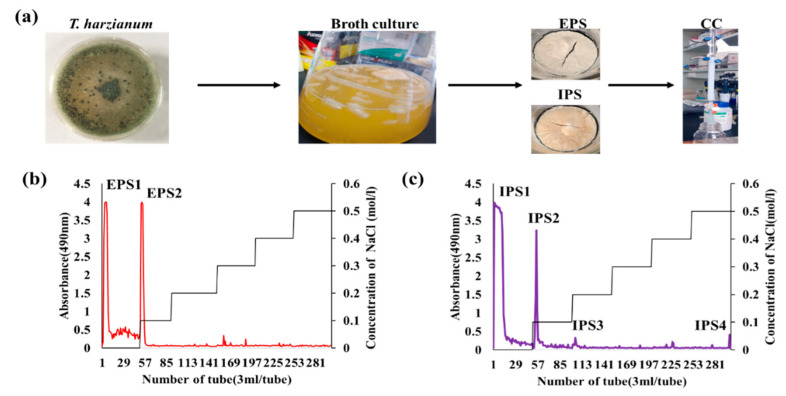
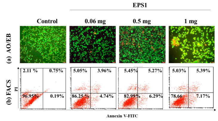
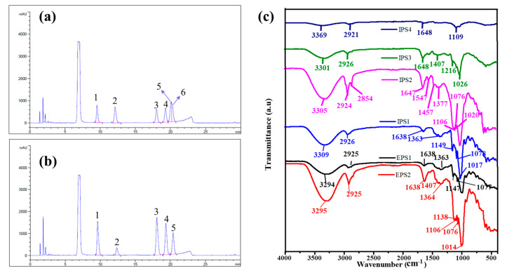
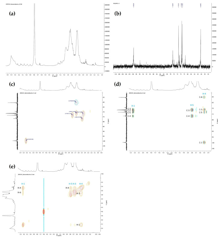
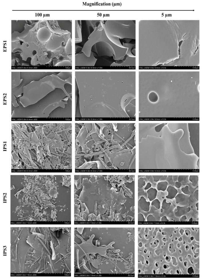
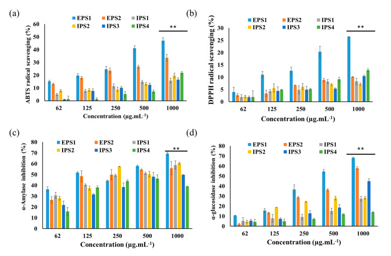
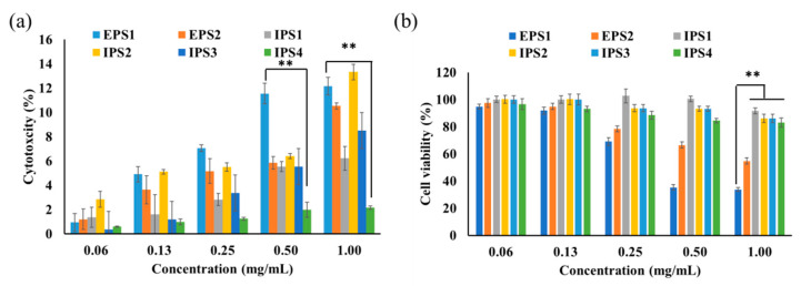
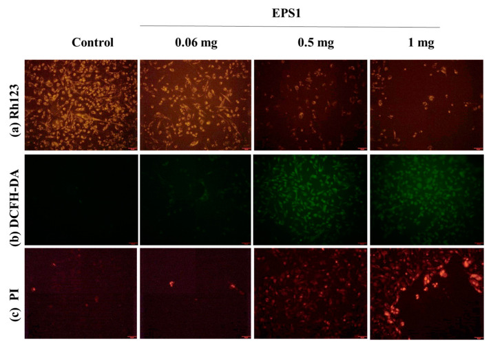
In this work, a total of six polysaccharides were isolated from culture filtrate (EPS1, EPS2) and mycelia (IPS1-IPS4) of . The HPLC analysis results showed that EPS1, EPS2, IPS1, and IPS2 were composed of mannose, ribose, glucose, galactose, and arabinose. The FT-IR, H, and C NMR chemical shifts confirmed that the signals in EPS1 mainly consist of (1→4)-linked α-d-glucopyranose. EPS1 and IPS1 showed a smooth and clean surface, while EPS2, IPS2, and IPS3 exhibited a microporous structure. Among polysaccharides, EPS1 displayed higher ABTS (47.09 ± 2.25% and DPPH (26.44 ± 0.12%) scavenging activities, as well as higher α-amylase (69.30 ± 1.28%) and α-glucosidase (68.22 ± 0.64%) inhibition activity than the other polysaccharides. EPS1 exhibited high cytotoxicity to MDA-MB293 cells, with an IC of 0.437 mg/mL, and this was also confirmed by cell staining and FACS assays. These results report the physicochemical and bioactive properties of polysaccharides from .
在这项工作中,从……的培养滤液(EPS1、EPS2)和菌丝体(IPS1 - IPS4)中总共分离出六种多糖。高效液相色谱分析结果表明,EPS1、EPS2、IPS1和IPS2由甘露糖、核糖、葡萄糖、半乳糖和阿拉伯糖组成。傅里叶变换红外光谱、氢谱和碳谱化学位移证实,EPS1中的信号主要由(1→4)连接的α - D - 吡喃葡萄糖组成。EPS1和IPS1呈现出光滑洁净的表面,而EPS2、IPS2和IPS3表现出微孔结构。在多糖中,EPS1表现出比其他多糖更高的ABTS清除活性(47.09 ± 2.25%)和DPPH清除活性(26.44 ± 0.12%),以及更高的α - 淀粉酶抑制活性(69.30 ± 1.28%)和α - 葡萄糖苷酶抑制活性(68.22 ± 0.64%)。EPS1对MDA - MB293细胞表现出高细胞毒性,IC50为0.437 mg/mL,细胞染色和流式细胞术分析也证实了这一点。这些结果报道了来自……的多糖的物理化学和生物活性特性。