Liu Junyan, Hormuth David A, Yang Jianchen, Yankeelov Thomas E
Department of Biomedical Engineering, The University of Texas at Austin, Austin, TX, United States.
Oden Institute for Computational Engineering and Sciences, The University of Texas at Austin, Austin, TX, United States.
Front Oncol. 2022 Feb 4;12:811415. doi: 10.3389/fonc.2022.811415. eCollection 2022.
Conventional radiobiology models, including the linear-quadratic model, do not explicitly account for the temporal effects of radiation, thereby making it difficult to make time-resolved predictions of tumor response to fractionated radiation. To overcome this limitation, we propose and validate an experimental-computational approach that predicts the changes in cell number over time in response to fractionated radiation.
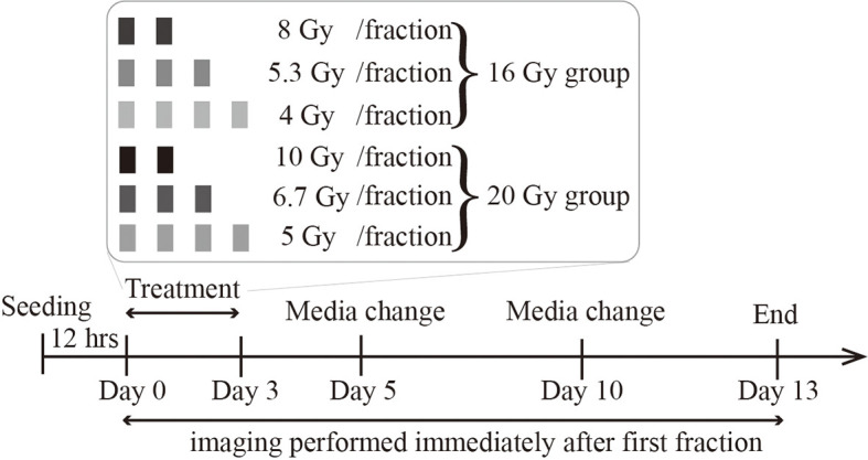
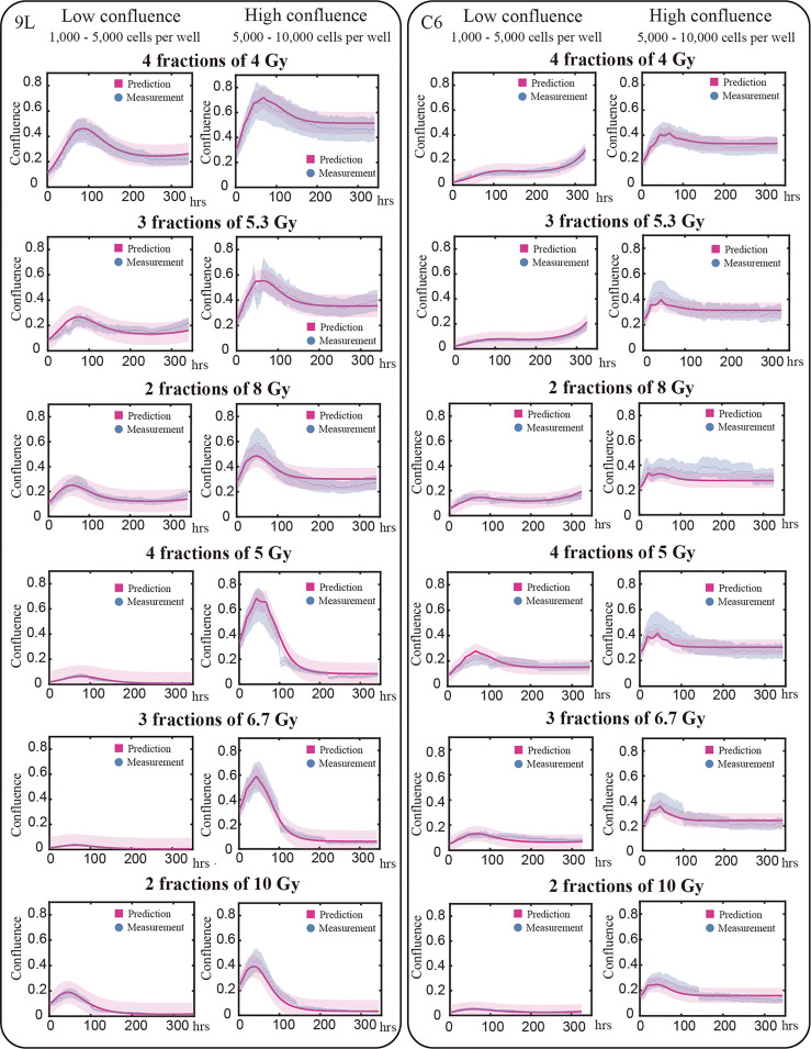
We irradiated 9L and C6 glioma cells with six different fractionation schemes yielding a total dose of either 16 Gy or 20 Gy, and then observed their response time-resolved microscopy. Phase-contrast images and Cytotox Red images (to label dead cells) were collected every 4 to 6 hours up to 330 hours post-radiation. Using 75% of the total data (i.e., 262 9L curves and 211 C6 curves), we calibrated a two-species model describing proliferative and senescent cells. We then applied the calibrated parameters to a validation dataset (the remaining 25% of the data, i.e., 91 9L curves and 74 C6 curves) to predict radiation response. Model predictions were compared to the microscopy measurements using the Pearson correlation coefficient (PCC) and the concordance correlation coefficient (CCC).
For the 9L cells, we observed PCCs and CCCs between the model predictions and validation data of (mean ± standard error) 0.96 ± 0.007 and 0.88 ± 0.013, respectively, across all fractionation schemes. For the C6 cells, we observed PCCs and CCCs between model predictions and the validation data were 0.89 ± 0.008 and 0.75 ± 0.017, respectively, across all fractionation schemes.
By proposing a time-resolved mathematical model of fractionated radiation response that can be experimentally verified , this study is the first to establish a framework for quantitative characterization and prediction of the dynamic radiobiological response of 9L and C6 gliomas to fractionated radiotherapy.
包括线性二次模型在内的传统放射生物学模型未明确考虑辐射的时间效应,因此难以对肿瘤对分割放疗的反应进行时间分辨预测。为克服这一局限性,我们提出并验证了一种实验 - 计算方法,该方法可预测细胞数量随时间的变化,以响应分割放疗。
我们用六种不同的分割方案对9L和C6胶质瘤细胞进行照射,总剂量分别为16 Gy或20 Gy,然后通过时间分辨显微镜观察其反应。在辐射后长达330小时内,每4至6小时收集相差图像和细胞毒性红图像(用于标记死亡细胞)。使用全部数据的75%(即262条9L曲线和211条C6曲线),我们校准了一个描述增殖细胞和衰老细胞的双物种模型。然后,我们将校准后的参数应用于验证数据集(剩余25%的数据,即91条9L曲线和74条C6曲线)以预测辐射反应。使用皮尔逊相关系数(PCC)和一致性相关系数(CCC)将模型预测结果与显微镜测量结果进行比较。
对于9L细胞,在所有分割方案中,我们观察到模型预测与验证数据之间的PCC和CCC分别为(平均值±标准误差)0.96±0.007和0.88±0.013。对于C6细胞,在所有分割方案中,我们观察到模型预测与验证数据之间的PCC和CCC分别为0.89±0.008和0.75±0.017。
通过提出一种可通过实验验证的分割放疗反应的时间分辨数学模型,本研究首次建立了一个框架,用于定量表征和预测9L和C6胶质瘤对分割放疗的动态放射生物学反应。