Key Laboratory of Oral Diseases of Jiangsu Province, Stomatological Institute, Nanjing Medical University, 136 Hanzhong Road, Nanjing, 210029, Jiangsu, China.
Endodontic Department, School of Stomatology, Nanjing Medical University, 136 Hanzhong Road, Nanjing, 210029, Jiangsu, China.
J Transl Med. 2022 May 13;20(1):208. doi: 10.1186/s12967-022-03412-9.
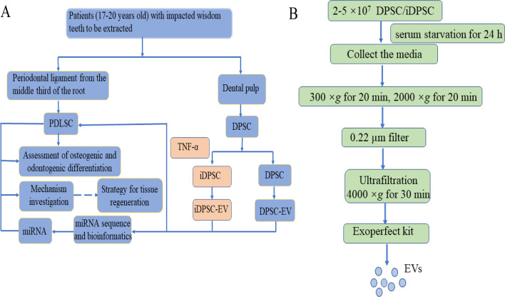
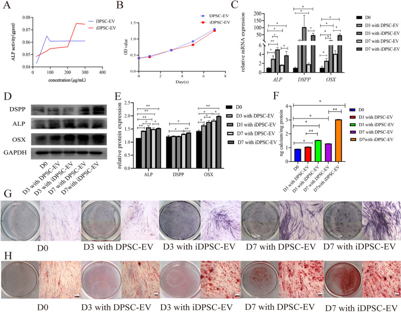
Extracellular vesicles (EVs) play a key role in constructing a microenvironment that favors the differentiation of stem cells. The present work aimed to determine the molecular mechanisms by which EV derived from inflammatory dental pulp stem cell (iDPSC-EV) influence periodontal ligament stem cells (PDLSCs) and provide a potential strategy for bone and dental pulp regeneration.
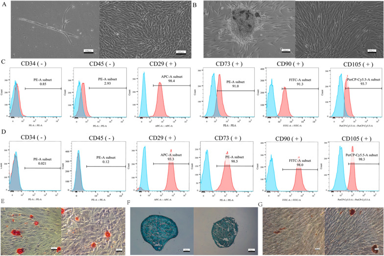
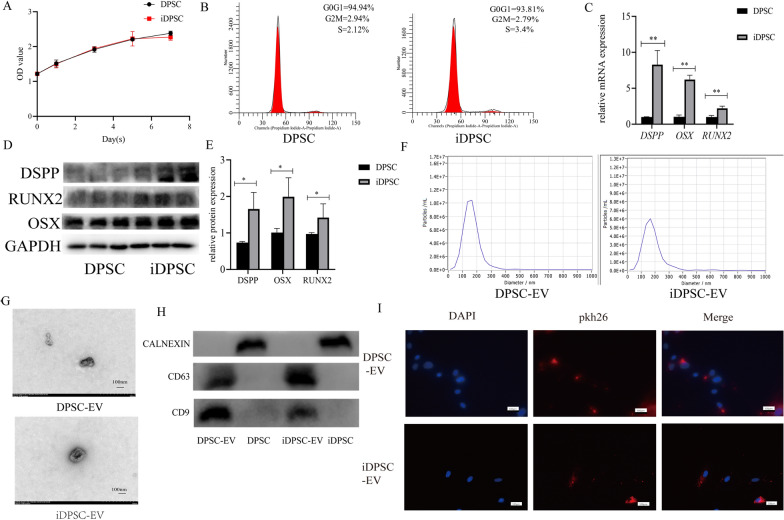
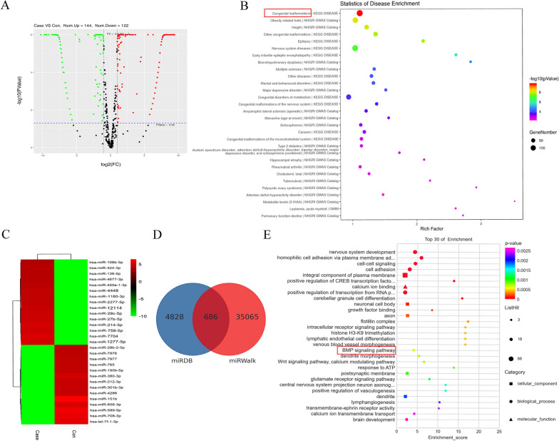
The osteogenic and odontogenic differentiation was assessed by quantitative real-time polymerase chain reaction (qRT-PCR), western blot, alkaline phosphatase (ALP) activity assay, ALP staining, alizarin red S (ARS) staining, and immunofluorescence staining. To detect proliferation, the Cell Counting Kit-8 (CCK-8) assay, and flow cytometry analysis were used. EVs were isolated by the Exoperfect kit and ultrafiltration and characterized by transmission electron microscopy (TEM), nanoparticle tracking analysis (NTA), and western blot. The expression profile of miRNAs in EVs was studied using miRNA sequence and bioinformatics, and one of the upregulated miRNAs was evaluated on PDLSCs.
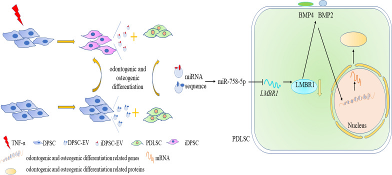
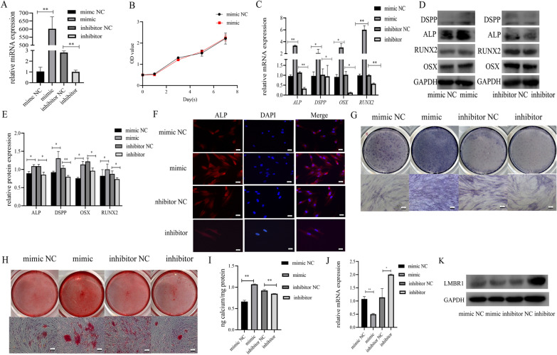
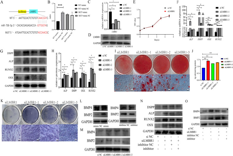
The inflammatory microenvironment stimulated osteogenic and odontogenic differentiation of DPSCs and iDPSC-EV behaved alike on PDLSCs. MiR-758-5p was upregulated in iDPSC-EV and was demonstrated to play a significant role in the osteogenic and odontogenic commitment of PDLSCs. A dual-luciferase reporter assay confirmed the binding site between miR-758-5p and limb development membrane protein 1 (LMBR1). The knockdown of LMBR1 also enhanced the above potential. Mechanically, bone morphogenetic protein (BMP) signaling was activated.
EVs from the inflammatory microenvironment enhanced the osteogenic and odontogenic differentiation of PDLSCs partly by shuttering LMBR1-targeting miR-758-5p via BMP signaling.
细胞外囊泡(EVs)在构建有利于干细胞分化的微环境中发挥关键作用。本研究旨在确定源自炎症牙髓干细胞(iDPSC-EV)的 EV 影响牙周膜干细胞(PDLSCs)的分子机制,并为骨和牙髓再生提供潜在策略。
通过定量实时聚合酶链反应(qRT-PCR)、western blot、碱性磷酸酶(ALP)活性测定、ALP 染色、茜素红 S(ARS)染色和免疫荧光染色评估成骨和成牙分化。使用细胞计数试剂盒-8(CCK-8)测定法和流式细胞术分析检测增殖。通过 Exoperfect 试剂盒和超滤分离 EVs,并通过透射电子显微镜(TEM)、纳米颗粒跟踪分析(NTA)和 western blot 进行表征。使用 miRNA 序列和生物信息学研究 EVs 中的 miRNA 表达谱,并评估其中一种上调的 miRNA 在 PDLSCs 上的作用。
炎症微环境刺激 DPSCs 的成骨和成牙分化,iDPSC-EV 对 PDLSCs 的作用与 DPSCs 相似。miR-758-5p 在 iDPSC-EV 中上调,并被证明在 PDLSCs 的成骨和成牙分化中发挥重要作用。双荧光素酶报告基因检测证实了 miR-758-5p 与肢体发育膜蛋白 1(LMBR1)之间的结合位点。LMBR1 的敲低也增强了上述潜能。机制上,骨形态发生蛋白(BMP)信号被激活。
炎症微环境来源的 EV 通过 BMP 信号抑制 LMBR1 靶向的 miR-758-5p,增强 PDLSCs 的成骨和成牙分化。