• 文献检索
  • 文档翻译
  • 深度研究
  • 学术资讯
  • Suppr Zotero 插件Zotero 插件
  • 邀请有礼
  • 套餐&价格
  • 历史记录
应用&插件
Suppr Zotero 插件Zotero 插件浏览器插件Mac 客户端Windows 客户端微信小程序
定价
高级版会员购买积分包购买API积分包
服务
文献检索文档翻译深度研究API 文档MCP 服务
关于我们
关于 Suppr公司介绍联系我们用户协议隐私条款
关注我们

Suppr 超能文献

核心技术专利:CN118964589B侵权必究
粤ICP备2023148730 号-1Suppr @ 2026

文献检索

告别复杂PubMed语法,用中文像聊天一样搜索,搜遍4000万医学文献。AI智能推荐,让科研检索更轻松。

立即免费搜索

文件翻译

保留排版,准确专业,支持PDF/Word/PPT等文件格式,支持 12+语言互译。

免费翻译文档

深度研究

AI帮你快速写综述,25分钟生成高质量综述,智能提取关键信息,辅助科研写作。

立即免费体验

毗邻性分析:用于多重组织成像数据的可视化空间邻域分析。

Visinity: Visual Spatial Neighborhood Analysis for Multiplexed Tissue Imaging Data.

出版信息

IEEE Trans Vis Comput Graph. 2023 Jan;29(1):106-116. doi: 10.1109/TVCG.2022.3209378. Epub 2022 Dec 16.

DOI:10.1109/TVCG.2022.3209378
PMID:36170403
原文链接:https://pmc.ncbi.nlm.nih.gov/articles/PMC10043053/
Abstract

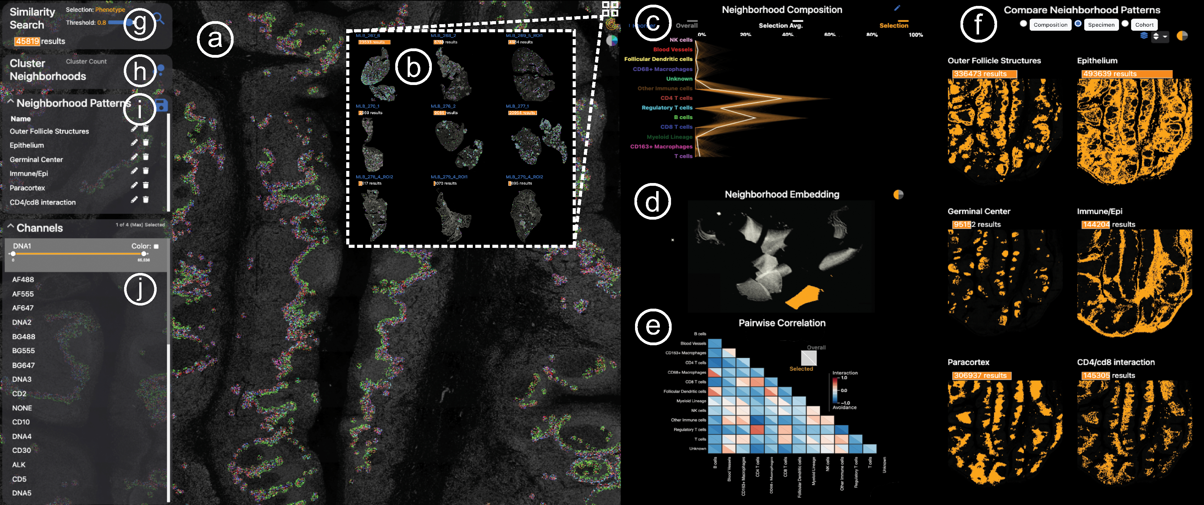
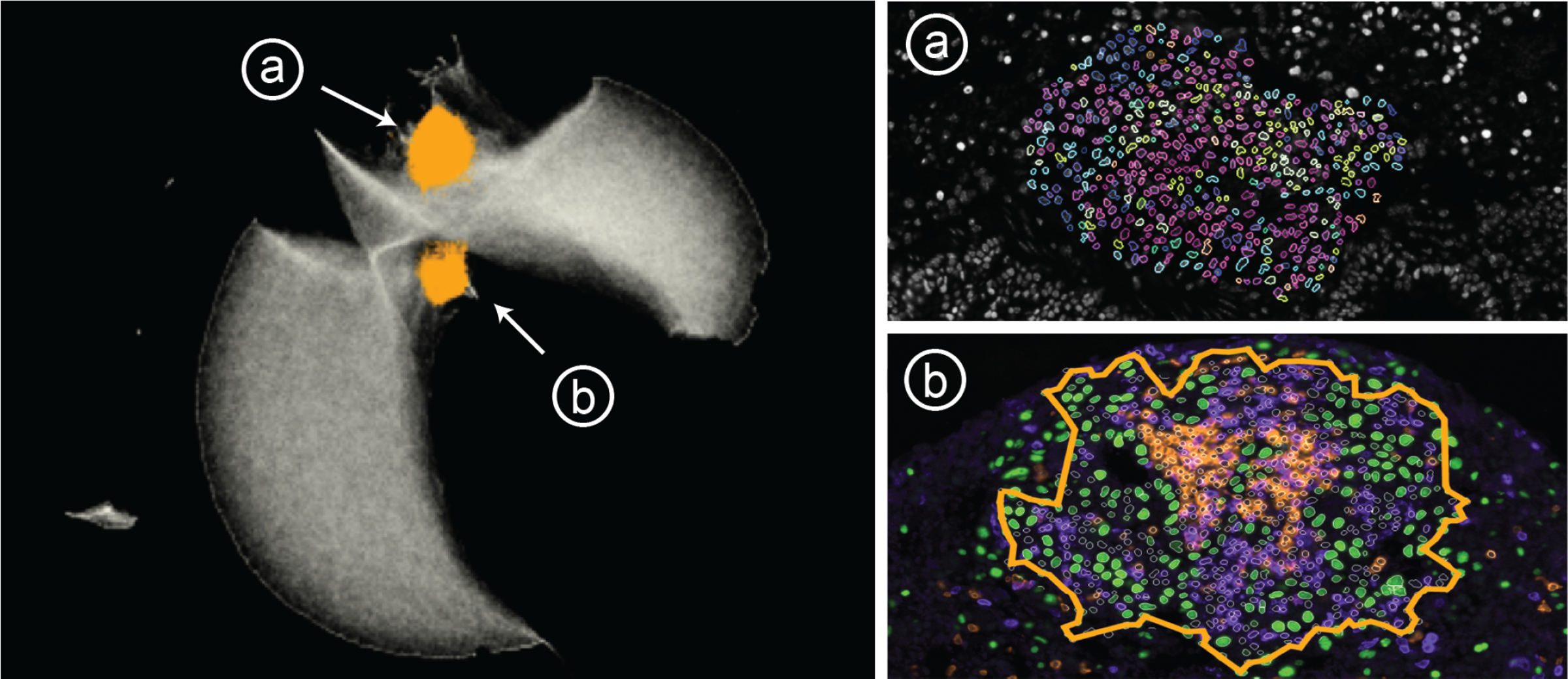
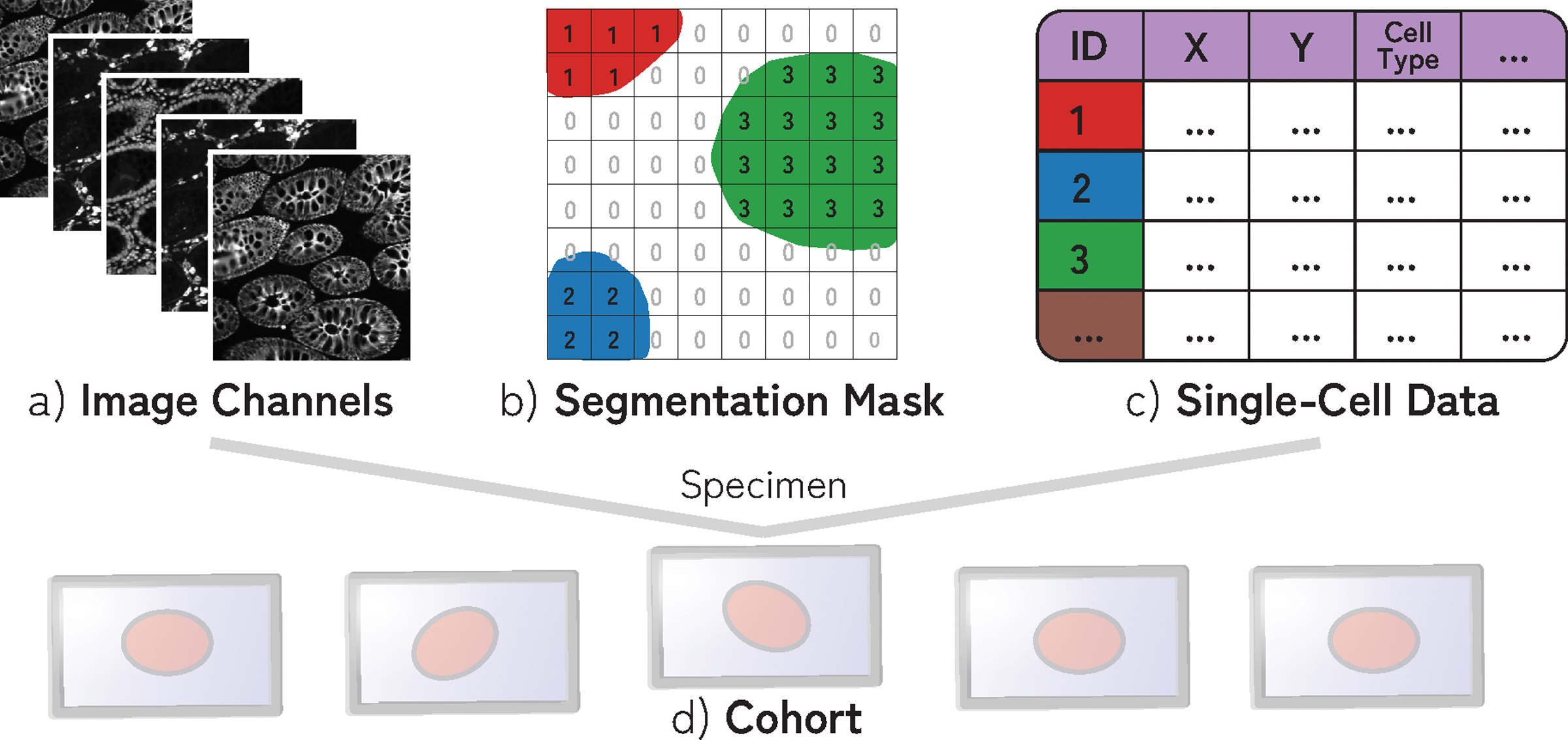
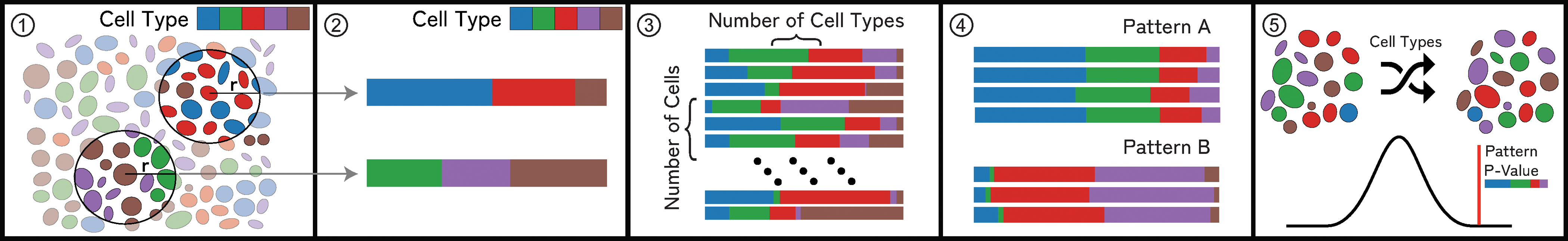
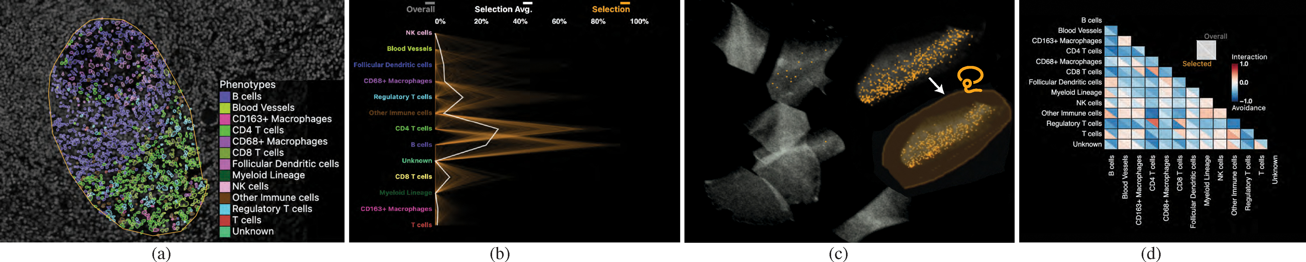
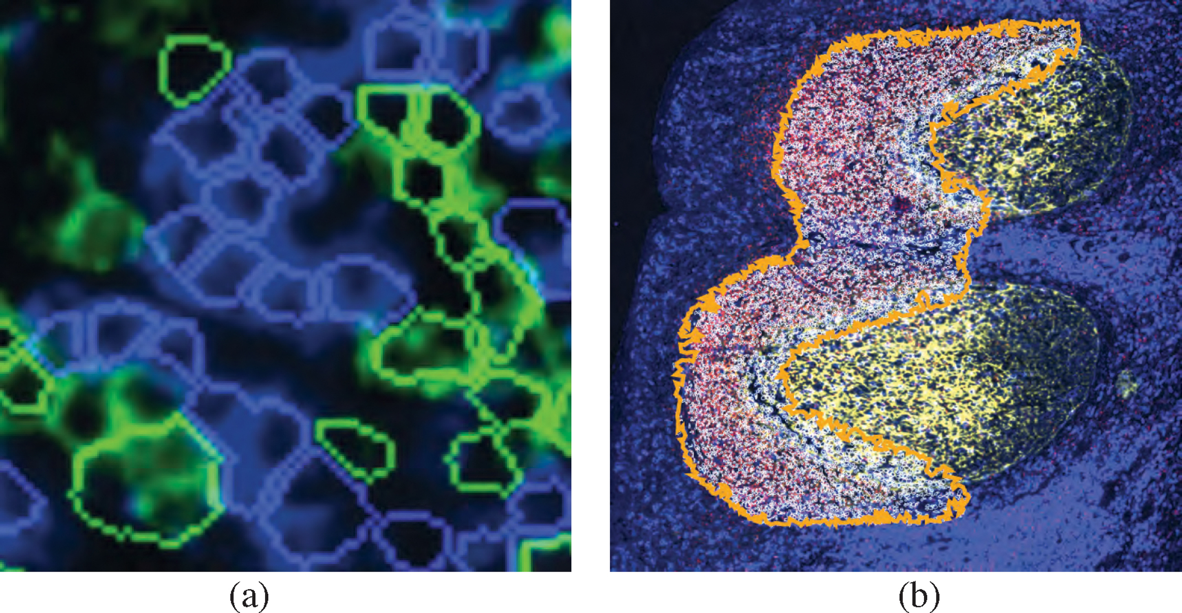
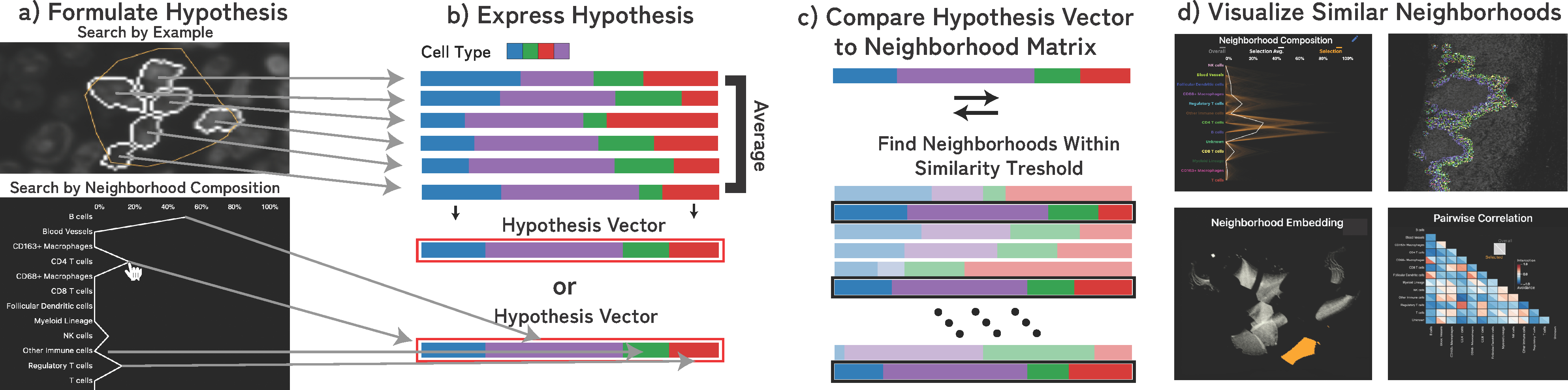
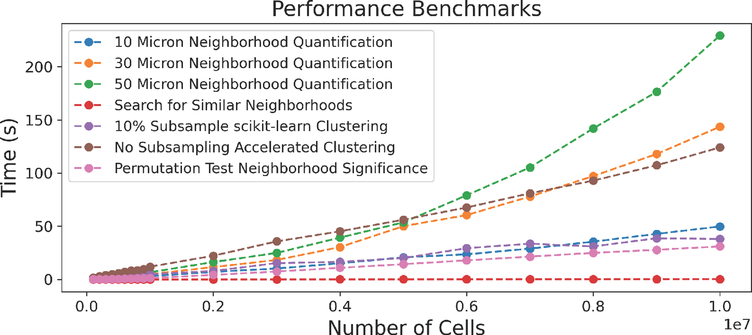
New highly-multiplexed imaging technologies have enabled the study of tissues in unprecedented detail. These methods are increasingly being applied to understand how cancer cells and immune response change during tumor development, progression, and metastasis, as well as following treatment. Yet, existing analysis approaches focus on investigating small tissue samples on a per-cell basis, not taking into account the spatial proximity of cells, which indicates cell-cell interaction and specific biological processes in the larger cancer microenvironment. We present Visinity, a scalable visual analytics system to analyze cell interaction patterns across cohorts of whole-slide multiplexed tissue images. Our approach is based on a fast regional neighborhood computation, leveraging unsupervised learning to quantify, compare, and group cells by their surrounding cellular neighborhood. These neighborhoods can be visually analyzed in an exploratory and confirmatory workflow. Users can explore spatial patterns present across tissues through a scalable image viewer and coordinated views highlighting the neighborhood composition and spatial arrangements of cells. To verify or refine existing hypotheses, users can query for specific patterns to determine their presence and statistical significance. Findings can be interactively annotated, ranked, and compared in the form of small multiples. In two case studies with biomedical experts, we demonstrate that Visinity can identify common biological processes within a human tonsil and uncover novel white-blood cell networks and immune-tumor interactions.

摘要

新的高度多重化成像技术使研究组织细节成为可能。这些方法越来越多地被用于了解癌细胞和免疫反应在肿瘤发展、进展和转移过程中以及治疗后的变化。然而,现有的分析方法侧重于逐个细胞地研究小的组织样本,而没有考虑到细胞的空间邻近性,这表明细胞间的相互作用和更大的癌症微环境中的特定生物学过程。我们提出了 Visinity,这是一种可扩展的可视化分析系统,用于分析整个幻灯片多重化组织图像队列中的细胞相互作用模式。我们的方法基于快速的区域邻域计算,利用无监督学习来量化、比较和根据其周围细胞邻域对细胞进行分组。这些邻域可以在探索性和验证性工作流程中进行可视化分析。用户可以通过可扩展的图像查看器和突出显示细胞邻域组成和空间排列的协调视图来探索组织中存在的空间模式。为了验证或完善现有的假设,用户可以查询特定模式以确定其存在和统计显著性。发现可以以小倍数的形式进行交互式注释、排名和比较。在与生物医学专家的两项案例研究中,我们证明了 Visinity 可以识别人扁桃体中的常见生物学过程,并揭示新的白细胞网络和免疫-肿瘤相互作用。

https://cdn.ncbi.nlm.nih.gov/pmc/blobs/f11b/10043053/89bb7ae6a548/nihms-1851534-f0011.jpg
https://cdn.ncbi.nlm.nih.gov/pmc/blobs/f11b/10043053/74b438f4ee67/nihms-1851534-f0001.jpg
https://cdn.ncbi.nlm.nih.gov/pmc/blobs/f11b/10043053/0c5b503cd063/nihms-1851534-f0002.jpg
https://cdn.ncbi.nlm.nih.gov/pmc/blobs/f11b/10043053/978bad582226/nihms-1851534-f0003.jpg
https://cdn.ncbi.nlm.nih.gov/pmc/blobs/f11b/10043053/1853471c1bf6/nihms-1851534-f0004.jpg
https://cdn.ncbi.nlm.nih.gov/pmc/blobs/f11b/10043053/0a5d6209b7db/nihms-1851534-f0005.jpg
https://cdn.ncbi.nlm.nih.gov/pmc/blobs/f11b/10043053/8abb76808662/nihms-1851534-f0006.jpg
https://cdn.ncbi.nlm.nih.gov/pmc/blobs/f11b/10043053/51df0e98a704/nihms-1851534-f0007.jpg
https://cdn.ncbi.nlm.nih.gov/pmc/blobs/f11b/10043053/85df1ed32192/nihms-1851534-f0008.jpg
https://cdn.ncbi.nlm.nih.gov/pmc/blobs/f11b/10043053/c2bbf5098f8f/nihms-1851534-f0009.jpg
https://cdn.ncbi.nlm.nih.gov/pmc/blobs/f11b/10043053/42b4dd1f5203/nihms-1851534-f0010.jpg
https://cdn.ncbi.nlm.nih.gov/pmc/blobs/f11b/10043053/89bb7ae6a548/nihms-1851534-f0011.jpg

相似文献

1
Visinity: Visual Spatial Neighborhood Analysis for Multiplexed Tissue Imaging Data.毗邻性分析:用于多重组织成像数据的可视化空间邻域分析。
IEEE Trans Vis Comput Graph. 2023 Jan;29(1):106-116. doi: 10.1109/TVCG.2022.3209378. Epub 2022 Dec 16.
2
Scope2Screen: Focus+Context Techniques for Pathology Tumor Assessment in Multivariate Image Data.Scope2Screen:用于多变量图像数据中病理肿瘤评估的聚焦+上下文技术。
IEEE Trans Vis Comput Graph. 2022 Jan;28(1):259-269. doi: 10.1109/TVCG.2021.3114786. Epub 2021 Dec 24.
3
Spatially resolved tissue imaging to analyze the tumor immune microenvironment: beyond cell-type densities.基于组织成像的肿瘤免疫微环境分析:超越细胞密度分析。
J Immunother Cancer. 2024 May 31;12(5):e008589. doi: 10.1136/jitc-2023-008589.
4
Facetto: Combining Unsupervised and Supervised Learning for Hierarchical Phenotype Analysis in Multi-Channel Image Data.Facetto:在多通道图像数据中结合无监督学习和监督学习进行分层表型分析
IEEE Trans Vis Comput Graph. 2020 Jan;26(1):227-237. doi: 10.1109/TVCG.2019.2934547. Epub 2019 Sep 10.
5
Synplex: In Silico Modeling of the Tumor Microenvironment From Multiplex Images.Synplex:从多重图像对肿瘤微环境进行计算建模。
IEEE Trans Med Imaging. 2023 Oct;42(10):3048-3058. doi: 10.1109/TMI.2023.3273950. Epub 2023 Oct 2.
6
Quantitative spatial evaluation of tumor-immune interactions in the immunotherapy setting of metastatic melanoma lymph nodes.转移性黑色素瘤淋巴结免疫治疗中肿瘤免疫相互作用的定量空间评估。
Front Immunol. 2022 Dec 5;13:1024039. doi: 10.3389/fimmu.2022.1024039. eCollection 2022.
7
NaroNet: Discovery of tumor microenvironment elements from highly multiplexed images.NaroNet:从高度多重化图像中发现肿瘤微环境元素。
Med Image Anal. 2022 May;78:102384. doi: 10.1016/j.media.2022.102384. Epub 2022 Feb 14.
8
Folic acid supplementation and malaria susceptibility and severity among people taking antifolate antimalarial drugs in endemic areas.在流行地区,服用抗叶酸抗疟药物的人群中,叶酸补充剂与疟疾易感性和严重程度的关系。
Cochrane Database Syst Rev. 2022 Feb 1;2(2022):CD014217. doi: 10.1002/14651858.CD014217.
9
Visual cohort comparison for spatial single-cell omics-data.空间单细胞组学数据的可视化队列比较
IEEE Trans Vis Comput Graph. 2021 Feb;27(2):733-743. doi: 10.1109/TVCG.2020.3030336. Epub 2021 Jan 28.
10
Semi-automated approaches for interrogating spatial heterogeneity of tissue samples.半自动化方法用于探究组织样本的空间异质性。
Sci Rep. 2024 Feb 29;14(1):5025. doi: 10.1038/s41598-024-55387-w.

引用本文的文献

1
Spatial Multiplexing and Omics.空间复用与组学
Nat Rev Methods Primers. 2024;4(1). doi: 10.1038/s43586-024-00330-6. Epub 2024 Aug 1.
2
CytoSpatio: Learning cell type spatial relationships using multirange, multitype point process models.细胞空间分析:使用多范围、多类型点过程模型学习细胞类型空间关系。
PLoS Comput Biol. 2025 Aug 21;21(8):e1013409. doi: 10.1371/journal.pcbi.1013409. eCollection 2025 Aug.
3
SEAL: Spatially-resolved Embedding Analysis with Linked Imaging Data.SEAL:基于链接成像数据的空间分辨嵌入分析
bioRxiv. 2025 Jul 28:2025.07.19.665696. doi: 10.1101/2025.07.19.665696.
4
Pathology-oriented multiplexing enables integrative disease mapping.面向病理学的多重分析可实现综合疾病图谱绘制。
Nature. 2025 Jul 18. doi: 10.1038/s41586-025-09225-2.
5
A toolkit for mapping cell identities in relation to neighbors reveals conserved patterning of neuromesodermal progenitor populations.一种用于绘制与相邻细胞相关的细胞身份图谱的工具包揭示了神经中胚层祖细胞群体的保守模式。
PLoS Biol. 2025 Jul 15;23(7):e3003244. doi: 10.1371/journal.pbio.3003244. eCollection 2025 Jul.
6
Advancing open-source visual analytics in digital pathology: A systematic review of tools, trends, and clinical applications.推进数字病理学中的开源视觉分析:对工具、趋势和临床应用的系统评价。
J Pathol Inform. 2025 May 23;18:100454. doi: 10.1016/j.jpi.2025.100454. eCollection 2025 Aug.
7
Spatial Patterning Analysis of Cellular Ensembles (SPACE) finds complex spatial organization at the cell and tissue levels.细胞集合体的空间模式分析(SPACE)在细胞和组织水平发现了复杂的空间组织。
Proc Natl Acad Sci U S A. 2025 Feb 11;122(6):e2412146122. doi: 10.1073/pnas.2412146122. Epub 2025 Feb 4.
8
Consensus tissue domain detection in spatial omics data using multiplex image labeling with regional morphology (MILWRM).利用具有区域形态学的多重图像标记(MILWRM)对空间组学数据中的共识组织域进行检测。
Commun Biol. 2024 Oct 30;7(1):1295. doi: 10.1038/s42003-024-06281-8.
9
Cell2Cell: Explorative Cell Interaction Analysis in Multi-Volumetric Tissue Data.细胞间:多体积组织数据中的探索性细胞相互作用分析
IEEE Trans Vis Comput Graph. 2025 Jan;31(1):569-579. doi: 10.1109/TVCG.2024.3456406. Epub 2024 Dec 3.
10
Aardvark: Composite Visualizations of Trees, Time-Series, and Images.土豚:树状图、时间序列和图像的复合可视化
IEEE Trans Vis Comput Graph. 2025 Jan;31(1):1290-1300. doi: 10.1109/TVCG.2024.3456193. Epub 2024 Nov 25.

本文引用的文献

1
Multiplexed 3D atlas of state transitions and immune interaction in colorectal cancer.多指标 3D 图谱描绘结直肠癌中状态转变和免疫相互作用。
Cell. 2023 Jan 19;186(2):363-381.e19. doi: 10.1016/j.cell.2022.12.028.
2
Viv: multiscale visualization of high-resolution multiplexed bioimaging data on the web.Viv:网络上高分辨率多路复用生物成像数据的多尺度可视化
Nat Methods. 2022 May;19(5):515-516. doi: 10.1038/s41592-022-01482-7.
3
The Spatial Landscape of Progression and Immunoediting in Primary Melanoma at Single-Cell Resolution.单细胞分辨率解析原发性黑色素瘤中的进展和免疫编辑的空间景观。
Cancer Discov. 2022 Jun 2;12(6):1518-1541. doi: 10.1158/2159-8290.CD-21-1357.
4
Spatial components of molecular tissue biology.分子组织生物学的空间组成部分。
Nat Biotechnol. 2022 Mar;40(3):308-318. doi: 10.1038/s41587-021-01182-1. Epub 2022 Feb 7.
5
MCMICRO: a scalable, modular image-processing pipeline for multiplexed tissue imaging.MCMICRO:一种用于多重组织成像的可扩展、模块化图像处理流水线。
Nat Methods. 2022 Mar;19(3):311-315. doi: 10.1038/s41592-021-01308-y. Epub 2021 Nov 25.
6
Cell segmentation in imaging-based spatial transcriptomics.基于成像的空间转录组学中的细胞分割
Nat Biotechnol. 2022 Mar;40(3):345-354. doi: 10.1038/s41587-021-01044-w. Epub 2021 Oct 14.
7
Scope2Screen: Focus+Context Techniques for Pathology Tumor Assessment in Multivariate Image Data.Scope2Screen:用于多变量图像数据中病理肿瘤评估的聚焦+上下文技术。
IEEE Trans Vis Comput Graph. 2022 Jan;28(1):259-269. doi: 10.1109/TVCG.2021.3114786. Epub 2021 Dec 24.
8
Peax: Interactive Visual Pattern Search in Sequential Data Using Unsupervised Deep Representation Learning.Peax:使用无监督深度表示学习在序列数据中进行交互式视觉模式搜索。
Comput Graph Forum. 2020 Jun;39(3):167-179. doi: 10.1111/cgf.13971. Epub 2020 Jul 18.
9
Cell segmentation-free inference of cell types from in situ transcriptomics data.无细胞分割的基于原位转录组学数据的细胞类型推断。
Nat Commun. 2021 Jun 10;12(1):3545. doi: 10.1038/s41467-021-23807-4.
10
Minerva: a light-weight, narrative image browser for multiplexed tissue images.密涅瓦:一种用于多重组织图像的轻量级叙事图像浏览器。
J Open Source Softw. 2020;5(54). doi: 10.21105/joss.02579. Epub 2020 Oct 15.