Elderdery Abozer Y, Alzahrani Badr, Hamza Siddiqa M A, Mostafa-Hedeab Gomaa, Mok Pooi Ling, Subbiah Suresh Kumar
Department of Clinical Laboratory Sciences, College of Applied Medical Sciences, Jouf University, Sakaka, Saudi Arabia.
Faculty of Medicine, Department of Pathology, Umm Alqura University Algunfuda, Mecca, Saudi Arabia.
Bioinorg Chem Appl. 2022 Sep 26;2022:1473922. doi: 10.1155/2022/1473922. eCollection 2022.
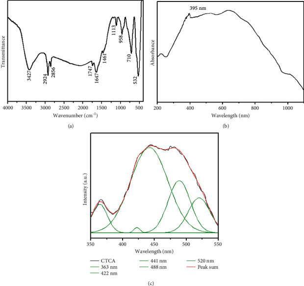
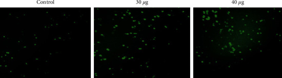
The main aim of this study was to synthesize copper oxide- (CuO-) titanium oxide- (TiO-) chitosan-amygdalin nanocomposites (CTCANc) and to characterize them physically and biologically (antimicrobial and anticancer activity using MOLT4 blood cancer cell line) to endorse their useful applications as potential drug candidates in anticancer avenues. CuO-TiO-chitosan-amygdalin nanocomposites were synthesized according to standard, reported methods. Physical characterization of the nanocomposites was performed using methods like X-ray diffractometer (XRD), and morphological and ultrastructural analysis of nanocomposites were done using electron microscope scanning and transmission. FTIR was recorded using a Perkin-Elmer spectrometer, and photoluminescence (PL) spectra were done using the spectrometer. Further, antibacterial activities were assessed using standard bacterial cultures. To demonstrate the nanocomposite's anticancer effects, MTT assay, morphological analysis, apoptosis studies using acridine orange/ethidium bromide (AO/EtBr) dual staining, reactive oxygen species (ROS) analysis, and levels of antioxidant enzymes were analyzed using the MOLT4 blood cancer cell line. Synthesized nanocomposites were characterized using XRD and showed various peaks, respectively, for CuO-TiO, amygdalin, and chitosan. MTT assay indicated an IC value of 38.41 g/ml concentration of CTCANc. Hence, 30 and 40 g/ml were used for the subsequent experiments. Morphological analysis, staining for apoptosis using AO/EtBr, mitochondrial membrane potential (MMP or ΔΨm) analysis, ROS analysis, and determination of the SOD, CAT, MDA, and GSH levels were performed. Observations like a significant loss of morphology, induction of apoptosis, elevated ROS, and decreased MMP were significant in 30 and 40 g/ml nanocomposite-treated cells when compared to control cells. The bimetallic nanocomposites exhibited typical nanocomposites characteristics and significant antibacterial and anticancer effects. The study results endorse the antibacterial, anticancer activity of CuO-TiO-chitosan-amygdalin nanocomposites and strongly suggest that further in-depth research using CuO-TiO-chitosan-amygdalin nanocomposites could reveal their efficacy in the clinical scenario.
本研究的主要目的是合成氧化铜-(CuO-)二氧化钛-(TiO-)壳聚糖-苦杏仁苷纳米复合材料(CTCANc),并对其进行物理和生物学表征(使用MOLT4血癌细胞系检测抗菌和抗癌活性),以认可其作为抗癌途径中潜在候选药物的有用应用。CuO-TiO-壳聚糖-苦杏仁苷纳米复合材料是根据标准的、已报道的方法合成的。使用X射线衍射仪(XRD)等方法对纳米复合材料进行物理表征,使用电子显微镜扫描和透射对纳米复合材料进行形态和超微结构分析。使用珀金埃尔默光谱仪记录傅里叶变换红外光谱(FTIR),使用该光谱仪进行光致发光(PL)光谱分析。此外,使用标准细菌培养物评估抗菌活性。为了证明纳米复合材料的抗癌效果,使用MOLT4血癌细胞系分析MTT法、形态分析、使用吖啶橙/溴化乙锭(AO/EtBr)双重染色的凋亡研究、活性氧(ROS)分析以及抗氧化酶水平。合成的纳米复合材料经XRD表征,分别显示出CuO-TiO、苦杏仁苷和壳聚糖的各种峰。MTT法表明CTCANc浓度为38.41 g/ml时的半数抑制浓度(IC)值。因此,后续实验使用30和40 g/ml。进行了形态分析、使用AO/EtBr进行凋亡染色、线粒体膜电位(MMP或ΔΨm)分析、ROS分析以及超氧化物歧化酶(SOD)、过氧化氢酶(CAT)、丙二醛(MDA)和谷胱甘肽(GSH)水平的测定。与对照细胞相比,在30和40 g/ml纳米复合材料处理的细胞中,观察到形态的显著丧失、凋亡的诱导、ROS升高和MMP降低等现象。双金属纳米复合材料表现出典型的纳米复合材料特性以及显著的抗菌和抗癌效果。研究结果认可了CuO-TiO-壳聚糖-苦杏仁苷纳米复合材料的抗菌、抗癌活性,并强烈表明使用CuO-TiO-壳聚糖-苦杏仁苷纳米复合材料进行进一步深入研究可能会揭示其在临床情况下的疗效。