Department of Electrical, Computer and Biomedical Engineering, University of Pavia, 27100 Pavia, Italy.
Research Institute for Applied Microelectronics (IUMA), University of Las Palmas de Gran Canaria (ULPGC), 35001 Las Palmas de Gran Canaria, Spain.
Sensors (Basel). 2022 Sep 21;22(19):7139. doi: 10.3390/s22197139.
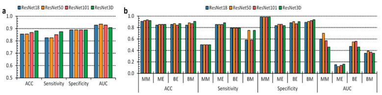
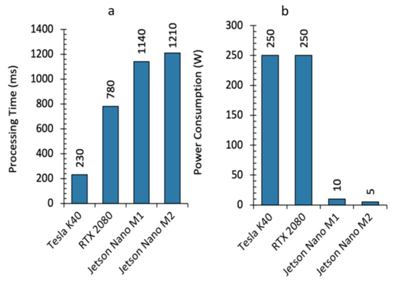
Cancer originates from the uncontrolled growth of healthy cells into a mass. Chromophores, such as hemoglobin and melanin, characterize skin spectral properties, allowing the classification of lesions into different etiologies. Hyperspectral imaging systems gather skin-reflected and transmitted light into several wavelength ranges of the electromagnetic spectrum, enabling potential skin-lesion differentiation through machine learning algorithms. Challenged by data availability and tiny inter and intra-tumoral variability, here we introduce a pipeline based on deep neural networks to diagnose hyperspectral skin cancer images, targeting a handheld device equipped with a low-power graphical processing unit for routine clinical testing. Enhanced by data augmentation, transfer learning, and hyperparameter tuning, the proposed architectures aim to meet and improve the well-known dermatologist-level detection performances concerning both benign-malignant and multiclass classification tasks, being able to diagnose hyperspectral data considering real-time constraints. Experiments show 87% sensitivity and 88% specificity for benign-malignant classification and specificity above 80% for the multiclass scenario. AUC measurements suggest classification performance improvement above 90% with adequate thresholding. Concerning binary segmentation, we measured skin DICE and IOU higher than 90%. We estimated 1.21 s, at most, consuming 5 Watts to segment the epidermal lesions with the U-Net++ architecture, meeting the imposed time limit. Hence, we can diagnose hyperspectral epidermal data assuming real-time constraints.
癌症起源于健康细胞的失控生长,形成一个肿块。生色团,如血红蛋白和黑色素,是皮肤光谱特性的特征,允许将病变分类为不同的病因。高光谱成像系统将皮肤反射和透射的光收集到电磁光谱的几个波长范围内,通过机器学习算法实现潜在的皮肤病变区分。由于数据可用性和微小的肿瘤内和肿瘤间变异性的挑战,我们在这里引入了一个基于深度神经网络的管道,用于诊断高光谱皮肤癌图像,针对的是配备低功耗图形处理单元的手持设备,用于常规临床测试。通过数据增强、迁移学习和超参数调整增强,所提出的架构旨在满足并提高著名皮肤科医生级别的检测性能,涉及良性-恶性和多类分类任务,能够考虑实时约束来诊断高光谱数据。实验表明,良性-恶性分类的敏感性为 87%,特异性为 88%,多类情况的特异性高于 80%。AUC 测量表明,适当的阈值下分类性能提高了 90%以上。关于二进制分割,我们测量的皮肤 DICE 和 IOU 高于 90%。使用 U-Net++架构分割表皮病变时,我们估计最多消耗 5 瓦,耗时 1.21 秒,满足规定的时间限制。因此,我们可以在假设实时约束的情况下诊断高光谱表皮数据。