Lee Yeong Chan, Cha Jiho, Shim Injeong, Park Woong-Yang, Kang Se Woong, Lim Dong Hui, Won Hong-Hee
Department of Digital Health, Samsung Advanced Institute for Health Sciences & Technology (SAIHST), Sungkyunkwan University, Samsung Medical Center, Seoul, Republic of Korea.
Research Institute for Future Medicine, Samsung Medical Center, Seoul, Republic of Korea.
NPJ Digit Med. 2023 Feb 2;6(1):14. doi: 10.1038/s41746-023-00748-4.
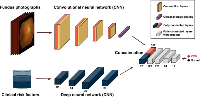
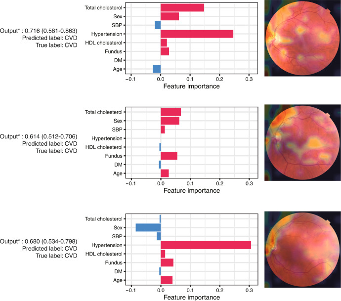
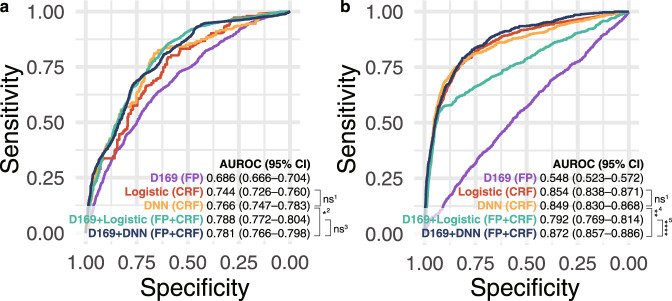
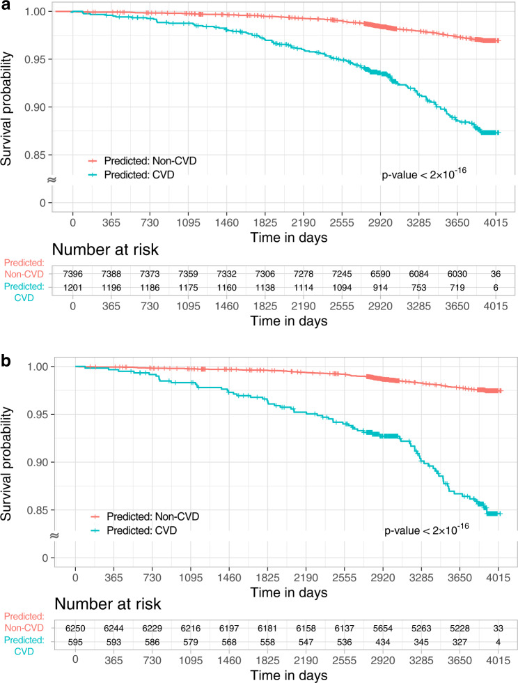
Cardiovascular disease (CVD), the leading cause of death globally, is associated with complicated underlying risk factors. We develop an artificial intelligence model to identify CVD using multimodal data, including clinical risk factors and fundus photographs from the Samsung Medical Center (SMC) for development and internal validation and from the UK Biobank for external validation. The multimodal model achieves an area under the receiver operating characteristic curve (AUROC) of 0.781 (95% confidence interval [CI] 0.766-0.798) in the SMC and 0.872 (95% CI 0.857-0.886) in the UK Biobank. We further observe a significant association between the incidence of CVD and the predicted risk from at-risk patients in the UK Biobank (hazard ratio [HR] 6.28, 95% CI 4.72-8.34). We visualize the importance of individual features in photography and traditional risk factors. The results highlight that non-invasive fundus photography can be a possible predictive marker for CVD.
心血管疾病(CVD)是全球主要死因,与复杂的潜在风险因素相关。我们开发了一种人工智能模型,使用多模态数据来识别CVD,这些数据包括来自三星医疗中心(SMC)用于模型开发和内部验证以及来自英国生物银行用于外部验证的临床风险因素和眼底照片。该多模态模型在SMC中的受试者操作特征曲线下面积(AUROC)为0.781(95%置信区间[CI]0.766 - 0.798),在英国生物银行中为0.872(95%CI 0.857 - 0.886)。我们进一步观察到在英国生物银行中,CVD发病率与高危患者的预测风险之间存在显著关联(风险比[HR]6.28,95%CI 4.72 - 8.34)。我们直观展示了摄影中的个体特征和传统风险因素的重要性。结果表明,非侵入性眼底摄影可能是CVD的一种预测标志物。