Microvision and Microdiagnostics Group (MVD), STI, Ecole Polytechnique Fédérale de Lausanne, Lausanne, Switzerland.
PLoS One. 2012;7(1):e30912. doi: 10.1371/journal.pone.0030912. Epub 2012 Jan 31.
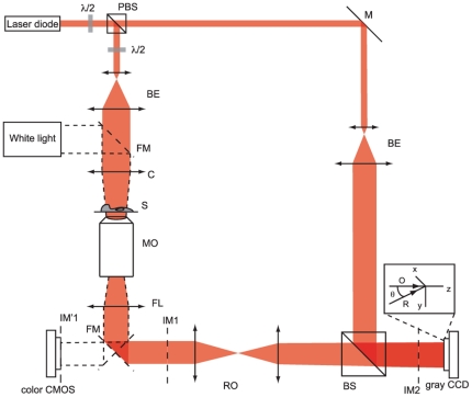
Digital holography provides a non-invasive measurement of the quantitative phase shifts induced by cells in culture, which can be related to cell volume changes. It has been shown previously that regulation of cell volume, in particular as it relates to ionic homeostasis, is crucially involved in the activation/inactivation of the cell death processes. We thus present here an application of digital holographic microscopy (DHM) dedicated to early and label-free detection of cell death.
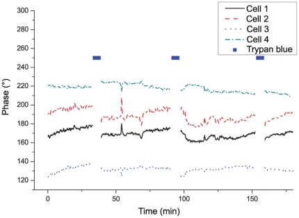
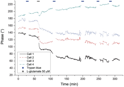
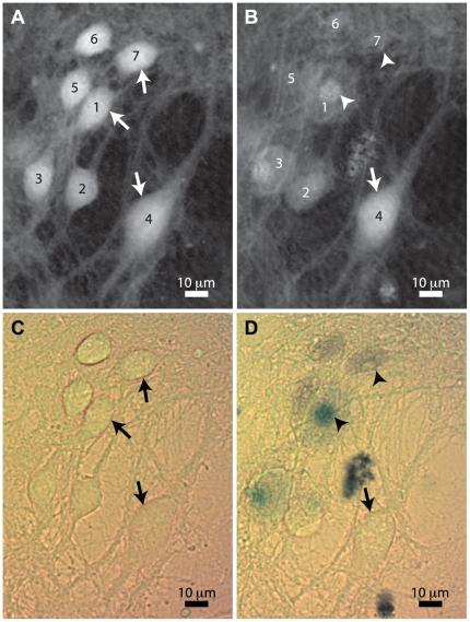
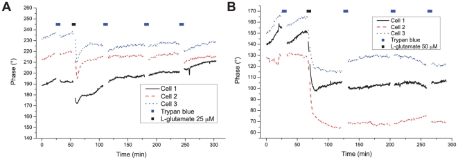
We provide quantitative measurements of phase signal obtained on mouse cortical neurons, and caused by early neuronal cell volume regulation triggered by excitotoxic concentrations of L-glutamate. We show that the efficiency of this early regulation of cell volume detected by DHM, is correlated with the occurrence of subsequent neuronal death assessed with the widely accepted trypan blue method for detection of cell viability.
The determination of the phase signal by DHM provides a simple and rapid optical method for the early detection of cell death.
数字全息术提供了一种非侵入式的测量方法,可以测量培养细胞中引起的定量相移,这与细胞体积变化有关。先前已经表明,细胞体积的调节,特别是与离子动态平衡有关的调节,对于细胞死亡过程的激活/失活至关重要。因此,我们在这里介绍了数字全息显微镜(DHM)在早期和无标记检测细胞死亡方面的应用。
我们提供了在小鼠皮质神经元上获得的相位信号的定量测量值,这些信号是由兴奋性谷氨酸盐浓度引发的早期神经元细胞体积调节引起的。我们表明,DHM 检测到的这种早期细胞体积调节的效率与随后用广泛接受的台盼蓝法检测细胞活力来评估的神经元死亡的发生相关。
DHM 通过确定相位信号,为早期检测细胞死亡提供了一种简单快速的光学方法。