• 文献检索
  • 文档翻译
  • 深度研究
  • 学术资讯
  • Suppr Zotero 插件Zotero 插件
  • 邀请有礼
  • 套餐&价格
  • 历史记录
应用&插件
Suppr Zotero 插件Zotero 插件浏览器插件Mac 客户端Windows 客户端微信小程序
定价
高级版会员购买积分包购买API积分包
服务
文献检索文档翻译深度研究API 文档MCP 服务
关于我们
关于 Suppr公司介绍联系我们用户协议隐私条款
关注我们

Suppr 超能文献

核心技术专利:CN118964589B侵权必究
粤ICP备2023148730 号-1Suppr @ 2026

文献检索

告别复杂PubMed语法,用中文像聊天一样搜索,搜遍4000万医学文献。AI智能推荐,让科研检索更轻松。

立即免费搜索

文件翻译

保留排版,准确专业,支持PDF/Word/PPT等文件格式,支持 12+语言互译。

免费翻译文档

深度研究

AI帮你快速写综述,25分钟生成高质量综述,智能提取关键信息,辅助科研写作。

立即免费体验

使用傅里叶和时域光学相干断层扫描技术对健康受试者视网膜神经纤维层厚度测量结果的比较。

Comparison of retinal nerve fiber layer thickness measurements in healthy subjects using fourier and time domain optical coherence tomography.

作者信息

Pinilla Isabel, Garcia-Martin Elena, Idoipe Miriam, Sancho Eva, Fuertes Isabel

机构信息

Ophthalmology Department, Lozano Blesa University Hospital, c/ San Juan Bosco 15, 50009 Zaragoza, Spain.

出版信息

J Ophthalmol. 2012;2012:107053. doi: 10.1155/2012/107053. Epub 2012 May 29.

DOI:10.1155/2012/107053
PMID:22690327
原文链接:https://pmc.ncbi.nlm.nih.gov/articles/PMC3368551/
Abstract

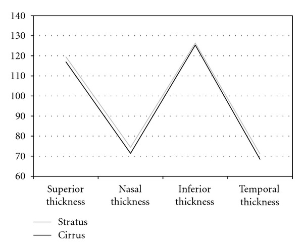
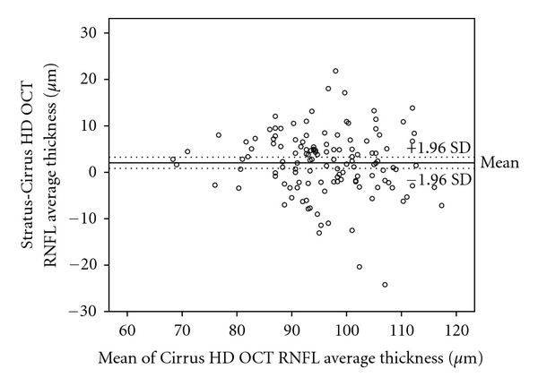
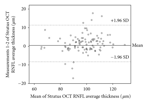
Purpose. To compare the retinal nerve fiber layer (RNFL) measurements using two different ocular coherence tomography (OCT) devices: Cirrus Fourier domain OCT and Stratus time domain OCT. To analyze reproducibility of Fourier domain measurements in healthy subjects. Methods. One hundred and thirty-two eyes of 132 healthy subjects were scaned on the same day with both instruments, separated by 10 minutes from each other. Thickness of quadrant, average and the 12 different areas around the optic nerve were compared between Cirrus and Stratus. Repeatability, intraclass correlation coefficients (ICCs), and coefficients of variation (COVs) were calculated in RNFL measurements provided by Fourier domain device. Results. The average thickness in the optic cube was 95.50 μm using Cirrus and 97.85 μm using Stratus. Average thickness and temporal quadrant showed significant differences using Cirrus and Stratus methods. Reproducibility was better with Fourier domain OCT (mean COV of 4.54%) than with Stratus time-domain OCT (mean COV of 5.57%). Conclusions. Both scan options give reproducible RNFL thickness measurement, but there are differences between them. Measurements obtained using Fourier domain device show better reproducibility.

摘要

目的。比较使用两种不同的光学相干断层扫描(OCT)设备测量视网膜神经纤维层(RNFL):Cirrus傅里叶域OCT和Stratus时域OCT。分析健康受试者傅里叶域测量的可重复性。方法。132名健康受试者的132只眼睛在同一天使用这两种仪器进行扫描,两次扫描间隔10分钟。比较Cirrus和Stratus之间象限、平均值以及视神经周围12个不同区域的厚度。计算傅里叶域设备提供的RNFL测量中的重复性、组内相关系数(ICC)和变异系数(COV)。结果。使用Cirrus时视盘立方体的平均厚度为95.50μm,使用Stratus时为97.85μm。使用Cirrus和Stratus方法时,平均厚度和颞侧象限显示出显著差异。傅里叶域OCT的可重复性(平均COV为4.54%)优于Stratus时域OCT(平均COV为5.57%)。结论。两种扫描方法均可获得可重复的RNFL厚度测量结果,但它们之间存在差异。使用傅里叶域设备获得的测量结果显示出更好的可重复性。

https://cdn.ncbi.nlm.nih.gov/pmc/blobs/52fc/3368551/10b4995a78f2/JOP2012-107053.004.jpg
https://cdn.ncbi.nlm.nih.gov/pmc/blobs/52fc/3368551/00149888ad1d/JOP2012-107053.001.jpg
https://cdn.ncbi.nlm.nih.gov/pmc/blobs/52fc/3368551/17c333fde5ae/JOP2012-107053.002.jpg
https://cdn.ncbi.nlm.nih.gov/pmc/blobs/52fc/3368551/c7121a5958ee/JOP2012-107053.003.jpg
https://cdn.ncbi.nlm.nih.gov/pmc/blobs/52fc/3368551/10b4995a78f2/JOP2012-107053.004.jpg

相似文献

1
Comparison of retinal nerve fiber layer thickness measurements in healthy subjects using fourier and time domain optical coherence tomography.使用傅里叶和时域光学相干断层扫描技术对健康受试者视网膜神经纤维层厚度测量结果的比较。
J Ophthalmol. 2012;2012:107053. doi: 10.1155/2012/107053. Epub 2012 May 29.
2
Macular and retinal nerve fiber layer thickness measurements in normal eyes with the Stratus OCT, the Cirrus HD-OCT, and the Topcon 3D OCT-1000.用 Stratus OCT、Cirrus HD-OCT 和 Topcon 3D OCT-1000 测量正常眼的黄斑和视网膜神经纤维层厚度。
J Glaucoma. 2011 Feb;20(2):118-25. doi: 10.1097/IJG.0b013e3181d786f8.
3
Comparison of retinal nerve fiber layer thickness measured by Cirrus HD and Stratus optical coherence tomography.Cirrus HD和Stratus光学相干断层扫描测量视网膜神经纤维层厚度的比较。
Ophthalmology. 2009 Jul;116(7):1264-70, 1270.e1. doi: 10.1016/j.ophtha.2008.12.045. Epub 2009 May 8.
4
Comparison of retinal nerve fiber layer measurements using time domain and spectral domain optical coherent tomography.使用时域和频域光学相干断层扫描技术测量视网膜神经纤维层的比较。
Ophthalmology. 2009 Jul;116(7):1271-7. doi: 10.1016/j.ophtha.2008.12.032. Epub 2009 Apr 22.
5
Comparison of retinal nerve fiber layer measured by time domain and spectral domain optical coherence tomography in optic neuritis.时域和频域光学相干断层扫描测量视神经炎患者视网膜神经纤维层的比较。
Eye (Lond). 2011 Feb;25(2):233-8. doi: 10.1038/eye.2010.206. Epub 2010 Dec 24.
6
Comparison of the diagnostic ability of retinal nerve fiber layer thickness measured using time domain and spectral domain optical coherence tomography in primary open angle glaucoma.时域和频域光学相干断层扫描测量视网膜神经纤维层厚度在原发性开角型青光眼中的诊断能力比较
Eye Sci. 2011 Sep;26(3):132-7, 142. doi: 10.3969/j.issn.1000-4432.2011.03.002.
7
Retinal nerve fiber layer imaging with spectral-domain optical coherence tomography: a variability and diagnostic performance study.光谱域光学相干断层扫描技术对视网膜神经纤维层的成像:变异性与诊断性能研究
Ophthalmology. 2009 Jul;116(7):1257-63, 1263.e1-2. doi: 10.1016/j.ophtha.2009.04.013. Epub 2009 May 22.
8
Fourier-domain OCT in multiple sclerosis patients: reproducibility and ability to detect retinal nerve fiber layer atrophy.傅里叶域光学相干断层扫描在多发性硬化症患者中的应用:重复性和检测视网膜神经纤维层萎缩的能力。
Invest Ophthalmol Vis Sci. 2011 Jun 13;52(7):4124-31. doi: 10.1167/iovs.10-6643.
9
Effects of changing operators and instruments on time-domain and spectral-domain OCT measurements of retinal nerve fiber layer thickness.更换操作人员和仪器对视网膜神经纤维层厚度的时域和频域光学相干断层扫描测量结果的影响。
Ophthalmic Surg Lasers Imaging. 2011 Jul-Aug;42(4):328-37. doi: 10.3928/15428877-20110603-05.
10
[Comparison of three optical coherence tomography devices, one time-domain and two fourier-domain, for the estimation of the retinal nerve fibre layer thickness].
Arch Soc Esp Oftalmol. 2010 May;85(5):165-73. doi: 10.1016/S0365-6691(10)55002-0.

引用本文的文献

1
Evaluation of Retinal Nerve Fiber Layer and Macular Thickness Pre- and Post-Chemotherapy With Carboplatin and Paclitaxel in Patients With Endometrial and Ovarian Cancer.子宫内膜癌和卵巢癌患者使用卡铂和紫杉醇化疗前后视网膜神经纤维层和黄斑厚度的评估
Cureus. 2023 Aug 22;15(8):e43943. doi: 10.7759/cureus.43943. eCollection 2023 Aug.
2
Retinal Nerve Fiber Layer Imaging with Two Different Spectral Domain Optical Coherence Tomographs: Normative Data for Romanian Children.两种不同光谱域光学相干断层扫描仪对视网膜神经纤维层的成像:罗马尼亚儿童的正常数据
Diagnostics (Basel). 2023 Apr 10;13(8):1377. doi: 10.3390/diagnostics13081377.
3

本文引用的文献

1
Optical coherence tomography in retinitis pigmentosa: reproducibility and capacity to detect macular and retinal nerve fiber layer thickness alterations.光学相干断层扫描在色素性视网膜炎中的应用:重复性及检测黄斑和视网膜神经纤维层厚度变化的能力。
Retina. 2012 Sep;32(8):1581-91. doi: 10.1097/IAE.0b013e318242b838.
2
Fourier-domain OCT in multiple sclerosis patients: reproducibility and ability to detect retinal nerve fiber layer atrophy.傅里叶域光学相干断层扫描在多发性硬化症患者中的应用:重复性和检测视网膜神经纤维层萎缩的能力。
Invest Ophthalmol Vis Sci. 2011 Jun 13;52(7):4124-31. doi: 10.1167/iovs.10-6643.
3
Intra and interoperator reproducibility of retinal nerve fibre and macular thickness measurements using Cirrus Fourier-domain OCT.
Optical Coherence Tomography in Patients with Alzheimer's Disease: What Can It Tell Us?
阿尔茨海默病患者的光学相干断层扫描:它能告诉我们什么?
Eye Brain. 2021 Jan 8;13:1-20. doi: 10.2147/EB.S235238. eCollection 2021.
4
Retinal nerve fibre layer thickness values and their associations with ocular and systemic parameters in Black South Africans.南非黑人的视网膜神经纤维层厚度值及其与眼部和全身参数的关联。
Afr Health Sci. 2016 Dec;16(4):1188-1194. doi: 10.4314/ahs.v16i4.39.
5
Structural and Functional Assessment in Patients Treated with Systemic Isotretinoin Using Optical Coherence Tomography and Frequency-Doubling Technology Perimetry.使用光学相干断层扫描和倍频技术视野检查法对接受系统性异维甲酸治疗的患者进行结构和功能评估。
Neuroophthalmology. 2013 May 31;37(3):100-103. doi: 10.3109/01658107.2013.792357. eCollection 2013.
6
Comparison of retinal nerve fiber layer and macular thickness measurements with Stratus OCT and OPKO/OTI OCT devices in healthy subjects.健康受试者中使用Stratus OCT和OPKO/OTI OCT设备测量视网膜神经纤维层和黄斑厚度的比较。
Int J Ophthalmol. 2015 Feb 18;8(1):98-103. doi: 10.3980/j.issn.2222-3959.2015.01.18. eCollection 2015.
7
Evaluation of retinal nerve fiber layer changes with oral isotretinoin treatment.评价口服异维 A 酸治疗对视网膜神经纤维层变化的影响。
Ophthalmol Ther. 2013 Jun;2(1):19-23. doi: 10.1007/s40123-012-0009-5. Epub 2013 Jan 26.
应用 Cirrus 傅里叶域光学相干断层扫描仪测量视网膜神经纤维和黄斑厚度的重复性(包括操作者内和操作者间)。
Acta Ophthalmol. 2011 Feb;89(1):e23-9. doi: 10.1111/j.1755-3768.2010.02045.x. Epub 2010 Nov 25.
4
Ultrahigh-resolution, high-speed, Fourier domain optical coherence tomography and methods for dispersion compensation.超高分辨率、高速傅里叶域光学相干断层扫描及色散补偿方法。
Opt Express. 2004 May 31;12(11):2404-22. doi: 10.1364/opex.12.002404.
5
Sensitivity advantage of swept source and Fourier domain optical coherence tomography.扫频光源和傅里叶域光学相干断层扫描的灵敏度优势。
Opt Express. 2003 Sep 8;11(18):2183-9. doi: 10.1364/oe.11.002183.
6
Signal strength is an important determinant of accuracy of nerve fiber layer thickness measurement by optical coherence tomography.信号强度是光学相干断层扫描测量神经纤维层厚度准确性的重要决定因素。
J Glaucoma. 2009 Mar;18(3):213-6. doi: 10.1097/IJG.0b013e31817eee20.
7
Comparison of Fourier-domain and time-domain optical coherence tomography in the detection of band atrophy of the optic nerve.傅里叶域与时域光学相干断层扫描技术在视神经带状萎缩检测中的比较
Am J Ophthalmol. 2009 Jan;147(1):56-63.e2. doi: 10.1016/j.ajo.2008.07.020. Epub 2008 Sep 6.
8
Retinal nerve fiber layer thickness in normal, ocular hypertensive, and glaucomatous Indian eyes: an optical coherence tomography study.正常、高眼压和青光眼印度人眼中视网膜神经纤维层厚度:光学相干断层扫描研究
J Glaucoma. 2008 Mar;17(2):122-7. doi: 10.1097/IJG.0b013e31814b9817.
9
Spectral-domain Cirrus high-definition optical coherence tomography is better than time-domain Stratus optical coherence tomography for evaluation of macular pathologic features in uveitis.在葡萄膜炎黄斑病变特征评估方面,频域Cirrus高清光学相干断层扫描比时域Stratus光学相干断层扫描更好。
Am J Ophthalmol. 2008 Jun;145(6):1018-1022. doi: 10.1016/j.ajo.2008.01.021. Epub 2008 Mar 17.
10
State-of-the-art retinal optical coherence tomography.视网膜光学相干断层扫描技术的最新进展。
Prog Retin Eye Res. 2008 Jan;27(1):45-88. doi: 10.1016/j.preteyeres.2007.07.005. Epub 2007 Aug 11.