Qiu Shangran, Joshi Prajakta S, Miller Matthew I, Xue Chonghua, Zhou Xiao, Karjadi Cody, Chang Gary H, Joshi Anant S, Dwyer Brigid, Zhu Shuhan, Kaku Michelle, Zhou Yan, Alderazi Yazan J, Swaminathan Arun, Kedar Sachin, Saint-Hilaire Marie-Helene, Auerbach Sanford H, Yuan Jing, Sartor E Alton, Au Rhoda, Kolachalama Vijaya B
Section of Computational Biomedicine, Department of Medicine, Boston University School of Medicine, Boston, MA, USA.
College of Arts and Sciences, Boston University, MA, USA.
Brain. 2020 Jun 1;143(6):1920-1933. doi: 10.1093/brain/awaa137.
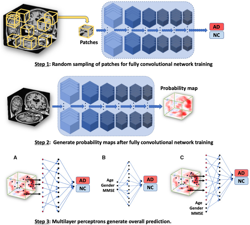
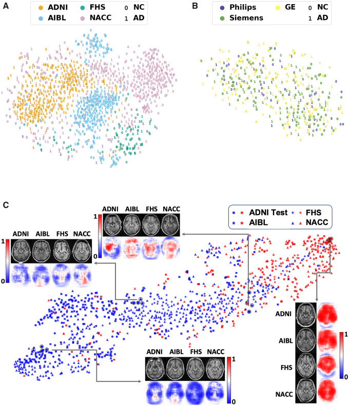
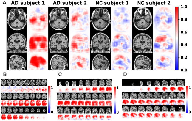
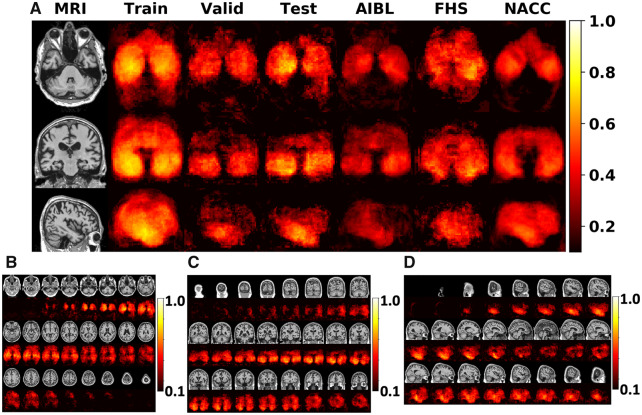
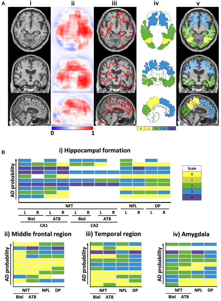
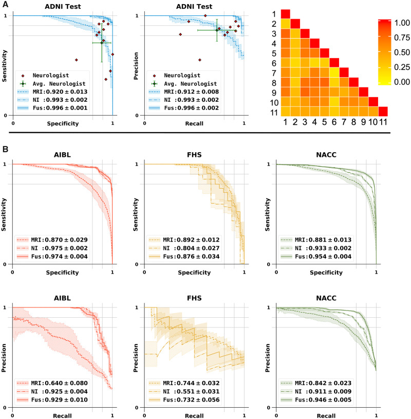
Alzheimer's disease is the primary cause of dementia worldwide, with an increasing morbidity burden that may outstrip diagnosis and management capacity as the population ages. Current methods integrate patient history, neuropsychological testing and MRI to identify likely cases, yet effective practices remain variably applied and lacking in sensitivity and specificity. Here we report an interpretable deep learning strategy that delineates unique Alzheimer's disease signatures from multimodal inputs of MRI, age, gender, and Mini-Mental State Examination score. Our framework linked a fully convolutional network, which constructs high resolution maps of disease probability from local brain structure to a multilayer perceptron and generates precise, intuitive visualization of individual Alzheimer's disease risk en route to accurate diagnosis. The model was trained using clinically diagnosed Alzheimer's disease and cognitively normal subjects from the Alzheimer's Disease Neuroimaging Initiative (ADNI) dataset (n = 417) and validated on three independent cohorts: the Australian Imaging, Biomarker and Lifestyle Flagship Study of Ageing (AIBL) (n = 382), the Framingham Heart Study (n = 102), and the National Alzheimer's Coordinating Center (NACC) (n = 582). Performance of the model that used the multimodal inputs was consistent across datasets, with mean area under curve values of 0.996, 0.974, 0.876 and 0.954 for the ADNI study, AIBL, Framingham Heart Study and NACC datasets, respectively. Moreover, our approach exceeded the diagnostic performance of a multi-institutional team of practicing neurologists (n = 11), and high-risk cerebral regions predicted by the model closely tracked post-mortem histopathological findings. This framework provides a clinically adaptable strategy for using routinely available imaging techniques such as MRI to generate nuanced neuroimaging signatures for Alzheimer's disease diagnosis, as well as a generalizable approach for linking deep learning to pathophysiological processes in human disease.
阿尔茨海默病是全球痴呆症的主要病因,随着人口老龄化,其发病负担不断增加,可能超过诊断和管理能力。目前的方法综合了患者病史、神经心理学测试和磁共振成像(MRI)来识别可能的病例,但有效做法的应用仍存在差异,且缺乏敏感性和特异性。在此,我们报告一种可解释的深度学习策略,该策略从MRI、年龄、性别和简易精神状态检查表得分的多模态输入中描绘出独特的阿尔茨海默病特征。我们的框架将一个全卷积网络与一个多层感知器相连接,前者从局部脑结构构建疾病概率的高分辨率图谱,并在准确诊断的过程中生成个体阿尔茨海默病风险的精确、直观可视化。该模型使用来自阿尔茨海默病神经影像倡议(ADNI)数据集(n = 417)的临床诊断为阿尔茨海默病的患者和认知正常的受试者进行训练,并在三个独立队列上进行验证:澳大利亚衰老成像、生物标志物和生活方式旗舰研究(AIBL)(n = 382)、弗雷明汉心脏研究(n = 102)和国家阿尔茨海默病协调中心(NACC)(n = 582)。使用多模态输入的模型在各数据集上的表现一致,ADNI研究、AIBL、弗雷明汉心脏研究和NACC数据集的曲线下面积均值分别为0.996、0.974、0.876和0.954。此外,我们的方法超过了一个由11名执业神经科医生组成的多机构团队的诊断性能,并且该模型预测的高风险脑区与死后组织病理学发现密切相关。这个框架提供了一种临床适用的策略,用于使用MRI等常规可用的成像技术来生成用于阿尔茨海默病诊断的细微神经影像特征,以及一种将深度学习与人类疾病病理生理过程相联系的通用方法。