Shu Tianyu, Zhang Yuchen, Sun Guo, Pan Yang, He Gang, Cheng Yilong, Li Ang, Pei Dandan
Key Laboratory of Shaanxi Province for Craniofacial Precision Medicine Research, College of Stomatology, Xi'an Jiaotong University, Xi'an, China.
State Key Laboratory of Military Stomatology, School of Stomatology, The Fourth Military Medical University, Xi'an, China.
Front Bioeng Biotechnol. 2021 Jan 7;8:621601. doi: 10.3389/fbioe.2020.621601. eCollection 2020.
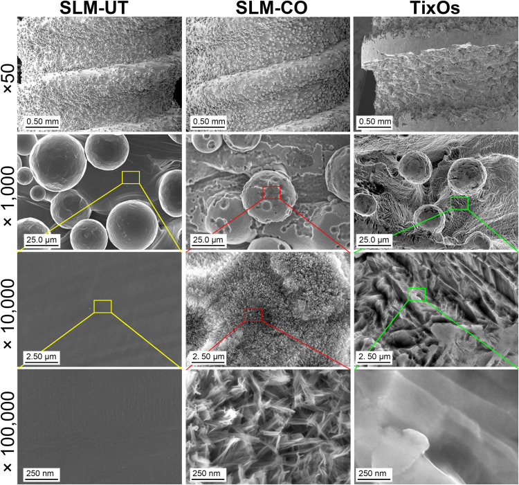
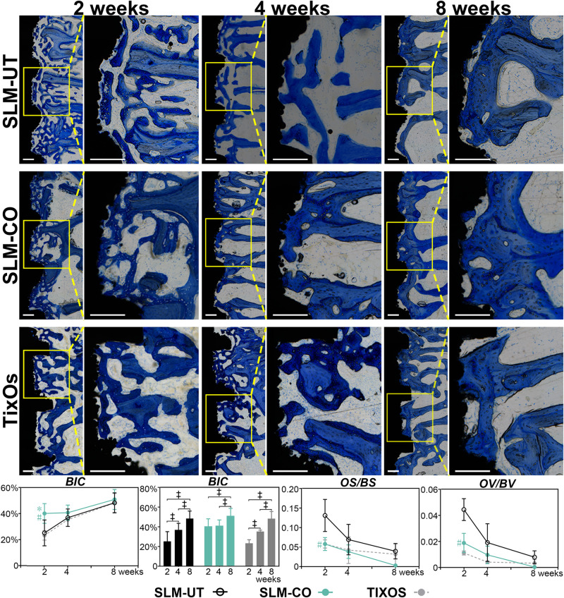
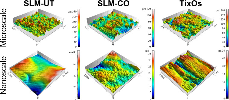
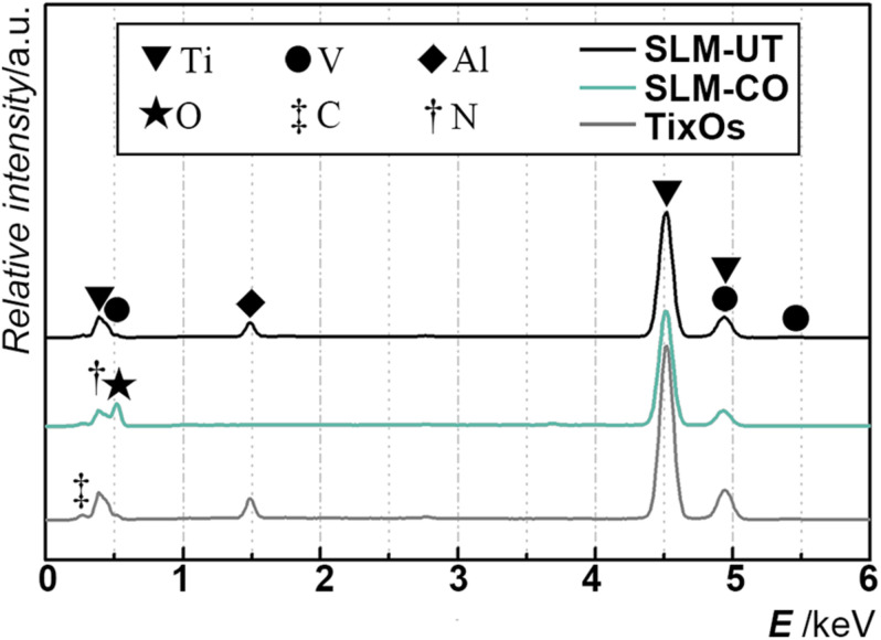
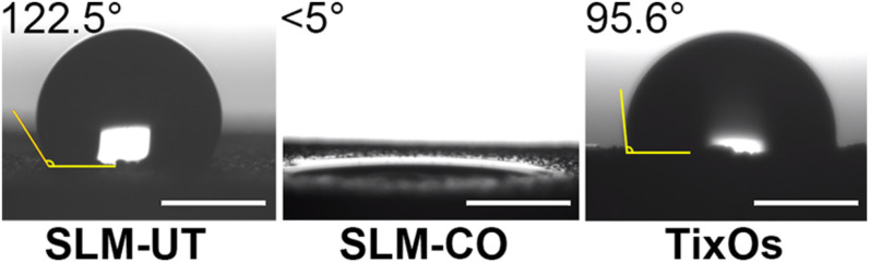
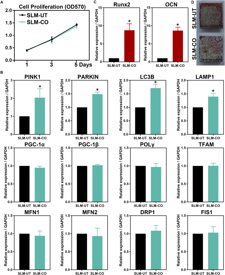
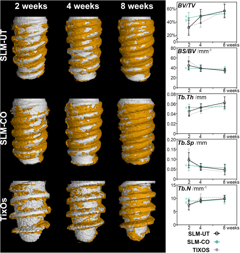
Currently, selective laser melting (SLM) has been thriving in implant dentistry for on-demand fabricating dental implants. Based on the coarse microtopography of SLM titanium surfaces, constructing nanostructure to form the hierarchical micro-nano topography is effective in enhancing osseointegration. Given that current nanomodification techniques of SLM implants, such as anodization and hydrothermal treatment, are facing the inadequacy in costly specific apparatus and reagents, there has been no recognized nanomodified SLM dental implants. The present study aimed to construct hierarchical micro-nano topography on self-made SLM dental implants by a simple and safe inorganic chemical oxidation, and to evaluate its contribution on osteoblastic cells bioactivity and osseointegration. The surface chemical and physical parameters were characterized by FE-SEM, EDS, profilometer, AFM, and contact angle meter. The alteration on bioactivity of MG-63 human osteoblastic cells were detected by qRT-PCR. Then the osseointegration was assessed by implanting implants on the femur condyle of New Zealand Rabbits. The hierarchical micro-nano topography was constituted by the microrough surface of SLM implants and nanoneedles (diameter: 20∼50 nm, height: 150∼250 nm), after nanomodifying SLM implants in 30% hydrogen peroxide and 30% hydrochloride acid (volume ratio 1:2.5) at room temperature for 36 h. Low chemical impurities content and high hydrophilicity were observed in the nanomodified group. Cell experiments on the nanomodified group showed higher expression of mitophagy related gene (PINK1, PARKIN, LC3B, and LAMP1) at 5 days and higher expression of osteogenesis related gene (Runx2 and OCN) at 14 days. In the early stage of bone formation, the nanomodified SLM implants demonstrated higher bone-to-implant contact. Intriguingly, the initial bone-to-implant contact of nanomodified SLM implants consisted of more mineralized bone with less immature osteoid. After the cessation of bone formation, the bone-to-implant contact of nanomodified SLM implants was equal to untreated SLM implants and marketable TixOs implants. The overall findings indicated that the inorganic chemical oxidized hierarchical micro-nano topography could enhance the bioactivity of osteoblastic cells, and consequently promote the peri-implant bone formation and mineralization of SLM dental implants. This study sheds some light on improvements in additive manufactured dental implants.
目前,选择性激光熔化(SLM)技术在口腔种植领域蓬勃发展,可按需制造牙科植入物。基于SLM钛表面粗糙的微观形貌,构建纳米结构以形成分级微纳形貌,对增强骨结合有效。鉴于当前SLM植入物的纳米改性技术,如阳极氧化和水热处理,面临着昂贵的特定设备和试剂不足的问题,目前尚无公认的纳米改性SLM牙科植入物。本研究旨在通过简单安全的无机化学氧化方法,在自制的SLM牙科植入物上构建分级微纳形貌,并评估其对成骨细胞生物活性和骨结合的作用。通过场发射扫描电子显微镜(FE-SEM)、能谱仪(EDS)、轮廓仪、原子力显微镜(AFM)和接触角测量仪对表面化学和物理参数进行表征。通过定量逆转录聚合酶链反应(qRT-PCR)检测MG-63人成骨细胞生物活性的变化。然后将植入物植入新西兰兔股骨髁,评估骨结合情况。在室温下将SLM植入物在30%过氧化氢和30%盐酸(体积比1:2.5)中纳米改性36小时后,分级微纳形貌由SLM植入物的微观粗糙表面和纳米针(直径:20∼50nm,高度:150∼250nm)组成。纳米改性组化学杂质含量低,亲水性高。纳米改性组的细胞实验显示,在第5天时与线粒体自噬相关基因(PINK1、PARKIN、LC3B和LAMP1)的表达较高,在第14天时与成骨相关基因(Runx2和OCN)的表达较高。在骨形成早期,纳米改性的SLM植入物表现出更高的骨-植入物接触率。有趣的是,纳米改性SLM植入物的初始骨-植入物接触由更多矿化骨和更少未成熟类骨质组成。骨形成停止后,纳米改性SLM植入物的骨-植入物接触率与未处理的SLM植入物和市售TixOs植入物相当。总体研究结果表明,无机化学氧化分级微纳形貌可增强成骨细胞的生物活性,从而促进SLM牙科植入物周围的骨形成和矿化。本研究为增材制造牙科植入物的改进提供了一些思路。