University of Tunis El Manar, Higher Institute of Medical Technologies of Tunis, Laboratory of Biophysics and Medical Technology, Tunis, Tunisia.
Université de Monastir - Laboratoire Technologie Imagerie Médicale - LTIM-LR12ES06, Faculté de Médecine de Monastir, 5019, Monastir, Tunisia; Université Paris-Est, Laboratoire d'Informatique Gaspard-Monge, Unité Mixte CNRS-UMLV-ESIEE UMR8049, ESIEE Paris Cité Descartes, BP99, 93162 Noisy Le Grand, France.
Clin Imaging. 2021 Aug;76:6-14. doi: 10.1016/j.clinimag.2021.01.019. Epub 2021 Jan 28.
SARS-CoV-2 is a worldwide health emergency with unrecognized clinical features. This paper aims to review the most recent medical imaging techniques used for the diagnosis of SARS-CoV-2 and their potential contributions to attenuate the pandemic. Recent researches, including artificial intelligence tools, will be described.
We review the main clinical features of SARS-CoV-2 revealed by different medical imaging techniques. First, we present the clinical findings of each technique. Then, we describe several artificial intelligence approaches introduced for the SARS-CoV-2 diagnosis.
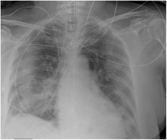
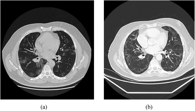
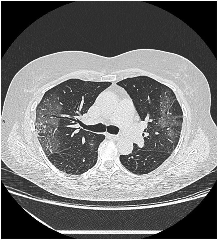
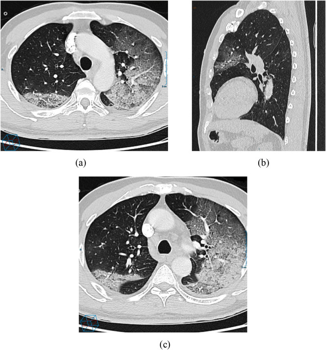
CT is the most accurate diagnostic modality of SARS-CoV-2. Additionally, ground-glass opacities and consolidation are the most common signs of SARS-CoV-2 in CT images. However, other findings such as reticular pattern, and crazy paving could be observed. We also found that pleural effusion and pneumothorax features are less common in SARS-CoV-2. According to the literature, the B lines artifacts and pleural line irregularities are the common signs of SARS-CoV-2 in ultrasound images. We have also stated the different studies, focusing on artificial intelligence tools, to evaluate the SARS-CoV-2 severity. We found that most of the reported works based on deep learning focused on the detection of SARS-CoV-2 from medical images while the challenge for the radiologists is how to differentiate between SARS-CoV-2 and other viral infections with the same clinical features.
The identification of SARS-CoV-2 manifestations on medical images is a key step in radiological workflow for the diagnosis of the virus and could be useful for researchers working on computer-aided diagnosis of pulmonary infections.
SARS-CoV-2 是一种具有未知临床特征的全球卫生紧急情况。本文旨在综述目前用于 SARS-CoV-2 诊断的最新医学影像学技术及其在减轻大流行方面的潜在贡献。将描述包括人工智能工具在内的最新研究。
我们回顾了不同医学影像学技术揭示的 SARS-CoV-2 的主要临床特征。首先,我们介绍了每种技术的临床发现。然后,我们描述了几种用于 SARS-CoV-2 诊断的人工智能方法。
CT 是 SARS-CoV-2 最准确的诊断方式。此外,磨玻璃影和实变是 CT 图像中 SARS-CoV-2 的最常见征象。然而,也可以观察到网状模式和铺路石征等其他征象。我们还发现胸腔积液和气胸特征在 SARS-CoV-2 中较少见。根据文献,B 线伪影和胸膜线不规则是 SARS-CoV-2 超声图像的常见征象。我们还提到了一些专注于人工智能工具的不同研究,以评估 SARS-CoV-2 的严重程度。我们发现,大多数基于深度学习的报道工作都集中在从医学图像中检测 SARS-CoV-2 上,而放射科医生的挑战是如何区分 SARS-CoV-2 和具有相同临床特征的其他病毒感染。
在放射科工作流程中,识别医学图像上的 SARS-CoV-2 表现是诊断该病毒的关键步骤,对于从事计算机辅助诊断肺部感染的研究人员可能很有用。