Department of Biosciences, Faculty of Science, Universiti Teknologi Malaysia, 81310, Johor Bahru, Johor, Malaysia.
IJN-UTM Cardiovascular Engineering Centre, Institute of Human Centered Engineering, Universiti Teknologi Malaysia, 81310, Johor Bahru, Johor, Malaysia.
Sci Rep. 2021 Mar 11;11(1):5634. doi: 10.1038/s41598-021-85149-x.
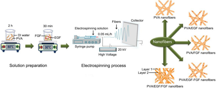
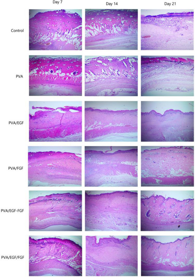
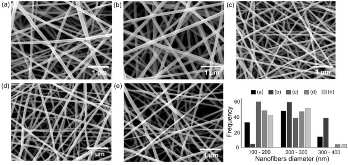
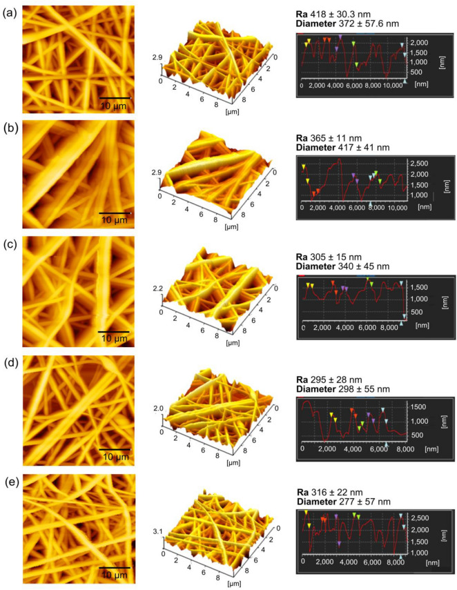
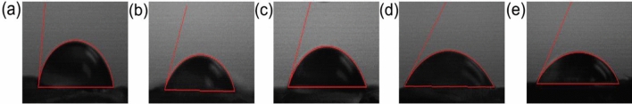
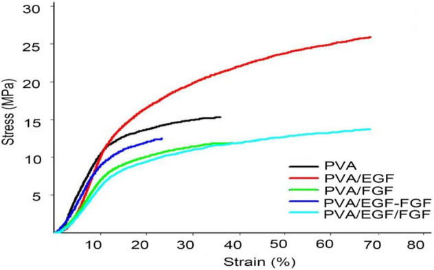
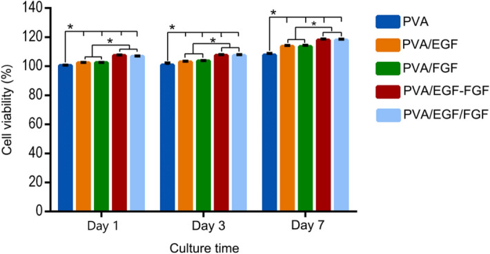
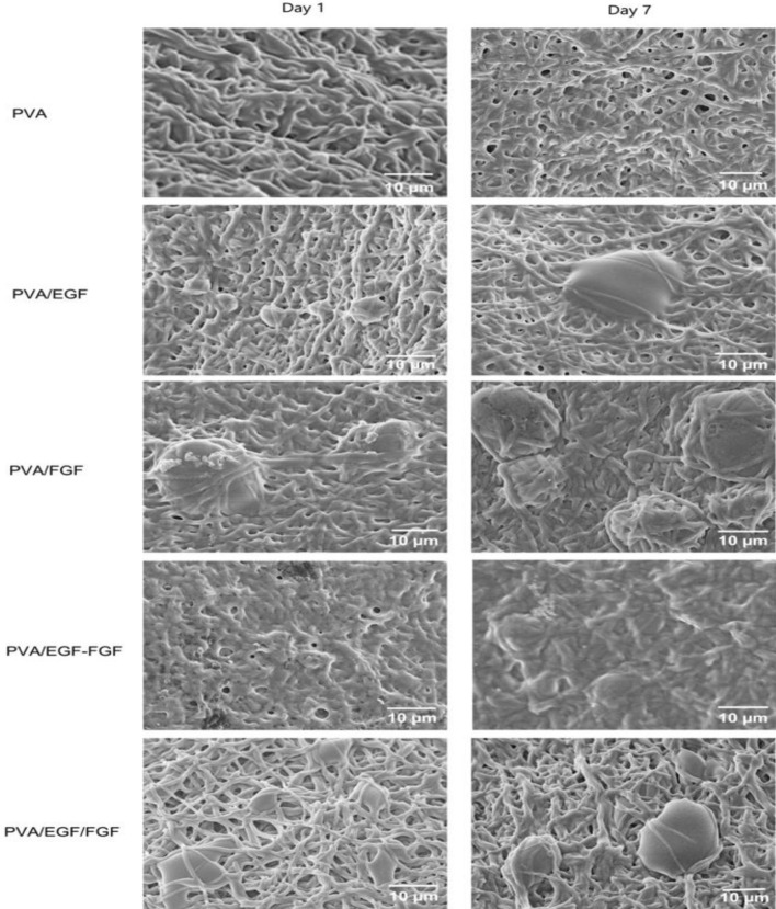
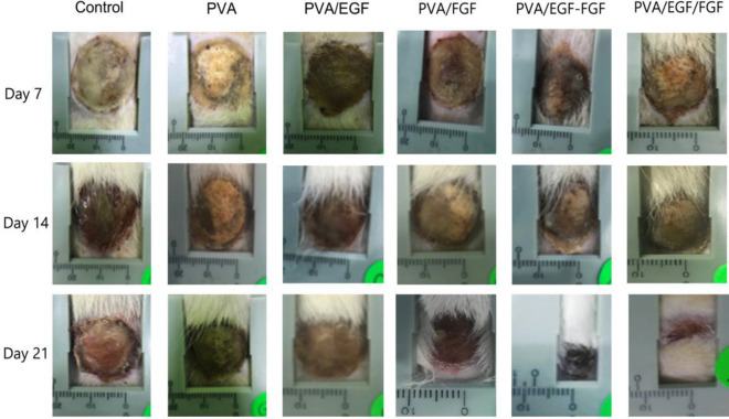
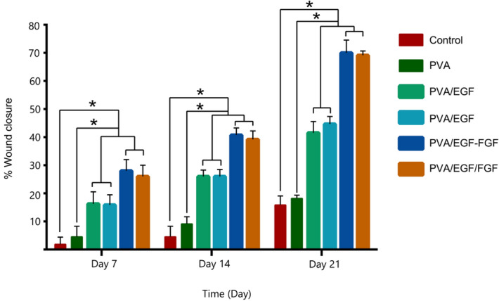
In this study, single, mix, multilayer Polyvinyl alcohol (PVA) electrospun nanofibers with epidermal growth factor (EGF) and fibroblast growth factor (FGF) were fabricated and characterized as a biological wound dressing scaffolds. The biological activities of the synthesized scaffolds have been verified by in vitro and in vivo studies. The chemical composition finding showed that the identified functional units within the produced nanofibers (O-H and N-H bonds) are attributed to both growth factors (GFs) in the PVA nanofiber membranes. Electrospun nanofibers' morphological features showed long protrusion and smooth morphology without beads and sprayed with an average range of 198-286 nm fiber diameter. The fiber diameters decrement and the improvement in wettability and surface roughness were recorded after GFs incorporated within the PVA Nanofibers, which indicated potential good adoption as biological dressing scaffolds due to the identified mechanical properties (Young's modulus) in between 18 and 20 MPa. The MTT assay indicated that the growth factor release from the PVA nanofibers has stimulated cell proliferation and promoted cell viability. In the cell attachment study, the GFs incorporated PVA nanofibers stimulated cell proliferation and adhered better than the PVA control sample and presented no cytotoxic effect. The in vivo studies showed that compared to the control and single PVA-GFs nanofiber, the mix and multilayer scaffolds gave a much more wound reduction at day 7 with better wound repair at day 14-21, which indicated to enhancing tissue regeneration, thus, could be a projected as a suitable burn wound dressing scaffold.
在这项研究中,制备了具有表皮生长因子(EGF)和成纤维细胞生长因子(FGF)的单一、混合、多层聚乙烯醇(PVA)电纺纳米纤维,并将其作为生物创伤敷料支架进行了表征。通过体外和体内研究验证了合成支架的生物活性。化学成分分析表明,在制备的纳米纤维中鉴定出的功能单元(O-H 和 N-H 键)归因于 PVA 纳米纤维膜中的两种生长因子(GFs)。电纺纳米纤维的形态特征显示出长突和光滑的形态,没有珠状物和喷涂,纤维直径平均范围为 198-286nm。在 GFs 掺入 PVA 纳米纤维后,记录到纤维直径减小,润湿性和表面粗糙度提高,这表明由于在 18 和 20MPa 之间确定的机械性能(杨氏模量),它们可能适合用作生物敷料支架。MTT 测定表明,PVA 纳米纤维中生长因子的释放刺激了细胞增殖并促进了细胞活力。在细胞附着研究中,掺入 GFs 的 PVA 纳米纤维刺激细胞增殖,比 PVA 对照样品更好地附着,并且没有细胞毒性作用。体内研究表明,与对照和单一 PVA-GFs 纳米纤维相比,混合和多层支架在第 7 天的伤口减少量更多,在第 14-21 天的伤口修复更好,这表明促进了组织再生,因此,可以作为一种合适的烧伤伤口敷料支架。