• 文献检索
  • 文档翻译
  • 深度研究
  • 学术资讯
  • Suppr Zotero 插件Zotero 插件
  • 邀请有礼
  • 套餐&价格
  • 历史记录
应用&插件
Suppr Zotero 插件Zotero 插件浏览器插件Mac 客户端Windows 客户端微信小程序
定价
高级版会员购买积分包购买API积分包
服务
文献检索文档翻译深度研究API 文档MCP 服务
关于我们
关于 Suppr公司介绍联系我们用户协议隐私条款
关注我们

Suppr 超能文献

核心技术专利:CN118964589B侵权必究
粤ICP备2023148730 号-1Suppr @ 2026

文献检索

告别复杂PubMed语法,用中文像聊天一样搜索,搜遍4000万医学文献。AI智能推荐,让科研检索更轻松。

立即免费搜索

文件翻译

保留排版,准确专业,支持PDF/Word/PPT等文件格式,支持 12+语言互译。

免费翻译文档

深度研究

AI帮你快速写综述,25分钟生成高质量综述,智能提取关键信息,辅助科研写作。

立即免费体验

基于图像引导的拉曼光谱探针跟踪技术用于肿瘤边界勾画。

Image-guided Raman spectroscopy probe-tracking for tumor margin delineation.

机构信息

Imperial College London, Department of Materials, London, United Kingdom.

Imperial College London, Department of Bioengineering, London, United Kingdom.

出版信息

J Biomed Opt. 2021 Mar;26(3). doi: 10.1117/1.JBO.26.3.036002.

DOI:10.1117/1.JBO.26.3.036002
PMID:33715315
原文链接:https://pmc.ncbi.nlm.nih.gov/articles/PMC7960531/
Abstract

SIGNIFICANCE

Tumor detection and margin delineation are essential for successful tumor resection. However, postsurgical positive margin rates remain high for many cancers. Raman spectroscopy has shown promise as a highly accurate clinical spectroscopic diagnostic modality, but its margin delineation capabilities are severely limited by the need for pointwise application.

AIM

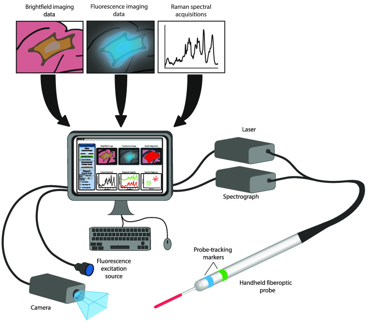
We aim to extend Raman spectroscopic diagnostics and develop a multimodal computer vision-based diagnostic system capable of both the detection and identification of suspicious lesions and the precise delineation of disease margins.

APPROACH

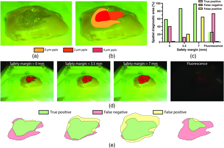
We first apply visual tracking of a Raman spectroscopic probe to achieve real-time tumor margin delineation. We then combine this system with protoporphyrin IX fluorescence imaging to achieve fluorescence-guided Raman spectroscopic margin delineation.

RESULTS

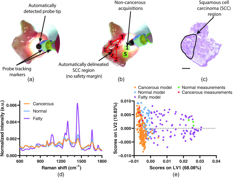
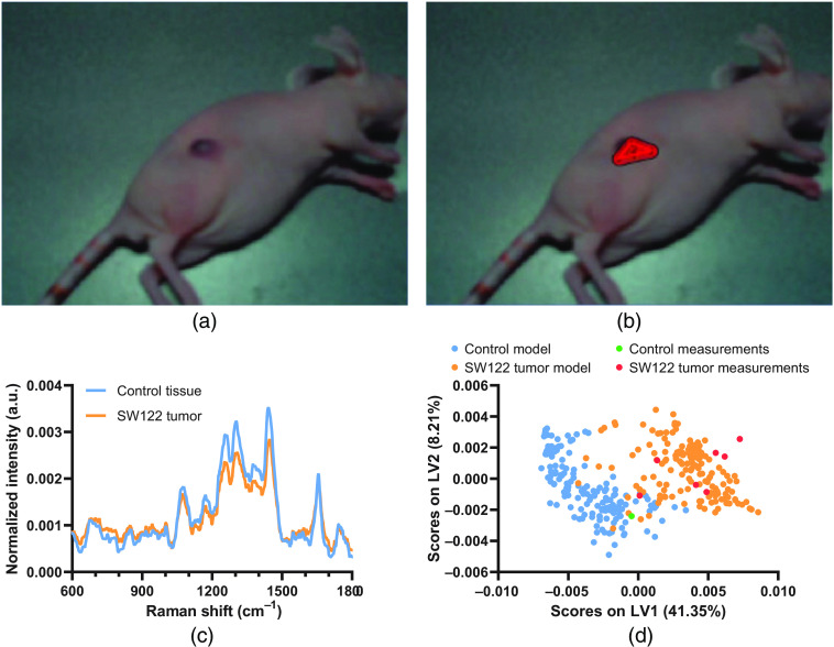
Our system enables real-time Raman spectroscopic tumor margin delineation for both ex vivo human tumor biopsies and an in vivo tumor xenograft mouse model. We then further demonstrate that the addition of protoporphyrin IX fluorescence imaging enables fluorescence-guided Raman spectroscopic margin delineation in a tissue phantom model.

CONCLUSIONS

Our image-guided Raman spectroscopic probe-tracking system enables tumor margin delineation and is compatible with both white light and fluorescence image guidance, demonstrating the potential for our system to be developed toward clinical tumor resection surgeries.

摘要

意义

肿瘤检测和边缘描绘对于成功进行肿瘤切除至关重要。然而,许多癌症的术后阳性边缘率仍然很高。拉曼光谱已被证明是一种非常准确的临床光谱诊断方式,但由于需要逐点应用,其边缘描绘能力受到严重限制。

目的

我们旨在扩展拉曼光谱诊断,并开发一种基于多模态计算机视觉的诊断系统,该系统能够同时检测和识别可疑病变,并精确描绘病变边缘。

方法

我们首先应用拉曼光谱探头的视觉跟踪来实现实时肿瘤边缘描绘。然后,我们将该系统与原卟啉 IX 荧光成像相结合,以实现荧光引导的拉曼光谱边缘描绘。

结果

我们的系统能够实时对离体人类肿瘤活检标本和体内肿瘤异种移植小鼠模型进行拉曼光谱肿瘤边缘描绘。然后,我们进一步证明,添加原卟啉 IX 荧光成像可以在组织体模模型中实现荧光引导的拉曼光谱边缘描绘。

结论

我们的图像引导拉曼光谱探头跟踪系统能够进行肿瘤边缘描绘,并且与白光和荧光图像引导兼容,这表明我们的系统有可能开发为临床肿瘤切除手术。

https://cdn.ncbi.nlm.nih.gov/pmc/blobs/5710/7960531/48965c16a10d/JBO-026-036002-g005.jpg
https://cdn.ncbi.nlm.nih.gov/pmc/blobs/5710/7960531/ad40843ca00a/JBO-026-036002-g001.jpg
https://cdn.ncbi.nlm.nih.gov/pmc/blobs/5710/7960531/67aa1a0fc311/JBO-026-036002-g002.jpg
https://cdn.ncbi.nlm.nih.gov/pmc/blobs/5710/7960531/8d7002e95b88/JBO-026-036002-g003.jpg
https://cdn.ncbi.nlm.nih.gov/pmc/blobs/5710/7960531/acddd9ae9888/JBO-026-036002-g004.jpg
https://cdn.ncbi.nlm.nih.gov/pmc/blobs/5710/7960531/48965c16a10d/JBO-026-036002-g005.jpg

相似文献

1
Image-guided Raman spectroscopy probe-tracking for tumor margin delineation.基于图像引导的拉曼光谱探针跟踪技术用于肿瘤边界勾画。
J Biomed Opt. 2021 Mar;26(3). doi: 10.1117/1.JBO.26.3.036002.
2
Surface-Enhanced Resonance Raman Scattering-Guided Brain Tumor Surgery Showing Prognostic Benefit in Rat Models.基于表面增强共振拉曼散射的脑肿瘤手术在大鼠模型中显示出预后获益。
ACS Appl Mater Interfaces. 2019 May 1;11(17):15241-15250. doi: 10.1021/acsami.9b00227. Epub 2019 Apr 17.
3
Pre-clinical evaluation of an image-guided in-situ Raman spectroscopy navigation system for targeted prostate cancer interventions.一种用于靶向前列腺癌介入的图像引导原位拉曼光谱导航系统的临床前评估。
Int J Comput Assist Radiol Surg. 2020 May;15(5):867-876. doi: 10.1007/s11548-020-02136-9. Epub 2020 Mar 29.
4
Enhancing surgical vision by using real-time imaging of αvβ3-integrin targeted near-infrared fluorescent agent.利用靶向 αvβ3 整合素的近红外荧光剂实时成像增强手术视野。
Ann Surg Oncol. 2011 Nov;18(12):3506-13. doi: 10.1245/s10434-011-1664-9. Epub 2011 Apr 21.
5
Delineating the tumor margin with intraoperative surface-enhanced Raman spectroscopy.术中表面增强拉曼光谱技术对肿瘤边界的描绘。
Anal Bioanal Chem. 2019 Jul;411(18):3993-4006. doi: 10.1007/s00216-019-01577-9. Epub 2019 Jan 25.
6
Real-time tracking of a diffuse reflectance spectroscopy probe used to aid histological validation of margin assessment in upper gastrointestinal cancer resection surgery.实时跟踪漫反射光谱探头,用于辅助上消化道癌症切除手术中边缘评估的组织学验证。
J Biomed Opt. 2022 Feb;27(2). doi: 10.1117/1.JBO.27.2.025001.
7
The complementary value of intraoperative fluorescence imaging and Raman spectroscopy for cancer surgery: combining the incompatibles.术中荧光成像和拉曼光谱在癌症手术中的互补价值:将不可兼容的技术结合起来。
Eur J Nucl Med Mol Imaging. 2022 Jun;49(7):2364-2376. doi: 10.1007/s00259-022-05705-z. Epub 2022 Feb 1.
8
Non-invasive Imaging of Cancer Using Surface-Enhanced Spatially Offset Raman Spectroscopy (SESORS).使用表面增强空间偏移拉曼光谱学(SESORS)进行癌症的无创成像。
Theranostics. 2019 Aug 13;9(20):5899-5913. doi: 10.7150/thno.36321. eCollection 2019.
9
Handheld macroscopic Raman spectroscopy imaging instrument for machine-learning-based molecular tissue margins characterization.基于机器学习的分子组织边界特征的手持式宏观拉曼光谱成像仪。
J Biomed Opt. 2021 Feb;26(2). doi: 10.1117/1.JBO.26.2.022911.
10
Raman spectroscopy for rapid intra-operative margin analysis of surgically excised tumour specimens.拉曼光谱法快速分析手术切除的肿瘤标本的术中边缘。
Analyst. 2019 Nov 4;144(22):6479-6496. doi: 10.1039/c9an01163c.

引用本文的文献

1
Raman analysis of lipids in cells: Current applications and future prospects.细胞中脂质的拉曼分析:当前应用与未来前景
J Pharm Anal. 2025 Apr;15(4):101136. doi: 10.1016/j.jpha.2024.101136. Epub 2024 Nov 1.
2
What's behind Margin Status in Oral Cancer?口腔癌切缘状态背后的因素是什么?
Indian J Otolaryngol Head Neck Surg. 2024 Oct;76(5):5001-5007. doi: 10.1007/s12070-024-04943-x. Epub 2024 Jul 30.
3
Pixel-level classification of pigmented skin cancer lesions using multispectral autofluorescence lifetime dermoscopy imaging.

本文引用的文献

1
Integrated photodynamic Raman theranostic system for cancer diagnosis, treatment, and post-treatment molecular monitoring.用于癌症诊断、治疗和治疗后分子监测的集成光动力拉曼治疗系统。
Theranostics. 2021 Jan 1;11(4):2006-2019. doi: 10.7150/thno.53031. eCollection 2021.
2
Raman spectroscopy for rapid intra-operative margin analysis of surgically excised tumour specimens.拉曼光谱法快速分析手术切除的肿瘤标本的术中边缘。
Analyst. 2019 Nov 4;144(22):6479-6496. doi: 10.1039/c9an01163c.
3
Fluorescence-guided surgery with aminolevulinic acid for low-grade gliomas.
使用多光谱自体荧光寿命皮肤镜成像对色素性皮肤癌病变进行像素级分类。
Biomed Opt Express. 2024 Jul 9;15(8):4557-4583. doi: 10.1364/BOE.523831. eCollection 2024 Aug 1.
4
Optical imaging technologies for cancer detection in low-resource settings.用于在资源匮乏地区进行癌症检测的光学成像技术。
Curr Opin Biomed Eng. 2023 Dec;28. doi: 10.1016/j.cobme.2023.100495. Epub 2023 Aug 23.
5
Point Projection Mapping System for Tracking, Registering, Labeling, and Validating Optical Tissue Measurements.用于跟踪、配准、标记和验证光学组织测量的点投影映射系统。
J Imaging. 2024 Jan 30;10(2):37. doi: 10.3390/jimaging10020037.
6
Intraoperative Assessment of Resection Margin in Oral Cancer: The Potential Role of Spectroscopy.口腔癌切除边缘的术中评估:光谱学的潜在作用。
Cancers (Basel). 2023 Dec 26;16(1):121. doi: 10.3390/cancers16010121.
7
Shedding Light on Colorectal Cancer: An In Vivo Raman Spectroscopy Approach Combined with Deep Learning Analysis.揭示结直肠癌的奥秘:体内拉曼光谱分析结合深度学习方法。
Int J Mol Sci. 2023 Nov 21;24(23):16582. doi: 10.3390/ijms242316582.
8
Combining electromyography and Raman spectroscopy: optical EMG.结合肌电图和拉曼光谱:光学 EMG。
Muscle Nerve. 2023 Oct;68(4):464-470. doi: 10.1002/mus.27937. Epub 2023 Jul 21.
9
Optical Tissue Phantoms for Quantitative Evaluation of Surgical Imaging Devices.用于手术成像设备定量评估的光学组织模拟体
Adv Photonics Res. 2023 Jan;4(1). doi: 10.1002/adpr.202200194. Epub 2022 Oct 13.
10
Hybrid confocal Raman endomicroscopy for morpho-chemical tissue characterization.用于形态化学组织表征的混合共焦拉曼内镜检查术。
Biomed Opt Express. 2022 Mar 21;13(4):2278-2285. doi: 10.1364/BOE.449110. eCollection 2022 Apr 1.
荧光引导手术联合氨基酮戊酸治疗低级别胶质瘤。
J Neurooncol. 2019 Jan;141(1):13-18. doi: 10.1007/s11060-018-03026-6. Epub 2018 Oct 26.
4
Optical Principles of Fluorescence-Guided Brain Tumor Surgery: A Practical Primer for the Neurosurgeon.荧光引导脑肿瘤手术的光学原理:神经外科医师实用入门
Neurosurgery. 2019 Sep 1;85(3):312-324. doi: 10.1093/neuros/nyy315.
5
Intra-operative spectroscopic assessment of surgical margins during breast conserving surgery.术中光谱评估在保乳手术中的手术切缘。
Breast Cancer Res. 2018 Jul 9;20(1):69. doi: 10.1186/s13058-018-1002-2.
6
Fluorescence-guided surgery of cancer: applications, tools and perspectives.荧光引导手术治疗癌症:应用、工具和前景。
Curr Opin Chem Biol. 2018 Aug;45:64-72. doi: 10.1016/j.cbpa.2018.03.002. Epub 2018 Mar 23.
7
Surgical oncology for gliomas: the state of the art.脑胶质瘤的外科治疗:现状。
Nat Rev Clin Oncol. 2018 Feb;15(2):112-125. doi: 10.1038/nrclinonc.2017.171. Epub 2017 Nov 21.
8
Light in diagnosis, therapy and surgery.光在诊断、治疗及手术中的应用。
Nat Biomed Eng. 2017;1. doi: 10.1038/s41551-016-0008. Epub 2017 Jan 10.
9
Real-time surgical tool tracking and pose estimation using a hybrid cylindrical marker.使用混合式圆柱标记进行实时手术工具跟踪和姿态估计。
Int J Comput Assist Radiol Surg. 2017 Jun;12(6):921-930. doi: 10.1007/s11548-017-1558-9. Epub 2017 Mar 24.
10
Fluorescence-guided resection of brain tumor: review of the significance of intraoperative quantification of protoporphyrin IX fluorescence.荧光引导下脑肿瘤切除术:术中原卟啉IX荧光定量的意义综述
Neurophotonics. 2017 Jan;4(1):011011. doi: 10.1117/1.NPh.4.1.011011. Epub 2017 Jan 12.