Gong Jing, Liu Jiyu, Li Haiming, Zhu Hui, Wang Tingting, Hu Tingdan, Li Menglei, Xia Xianwu, Hu Xianfang, Peng Weijun, Wang Shengping, Tong Tong, Gu Yajia
Department of Radiology, Fudan University Shanghai Cancer Center, 270 Dongan Road, Shanghai 200032, China.
Department of Oncology, Shanghai Medical College, Fudan University, Shanghai 200032, China.
Cancers (Basel). 2021 Jun 30;13(13):3300. doi: 10.3390/cancers13133300.
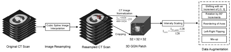
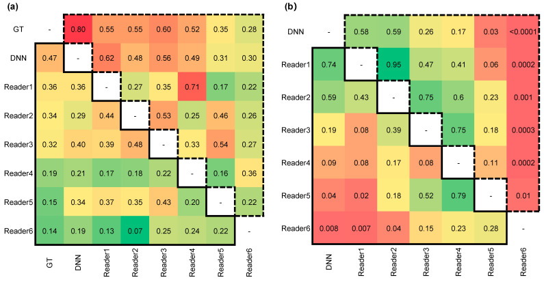
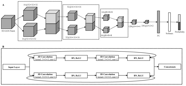
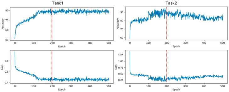
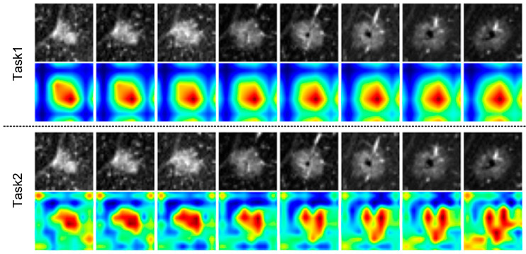
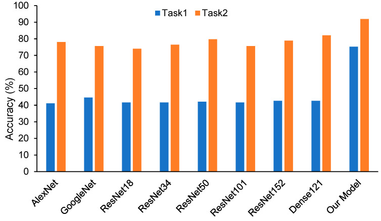
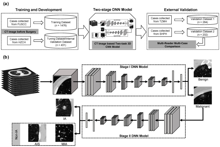
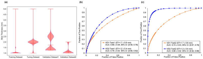
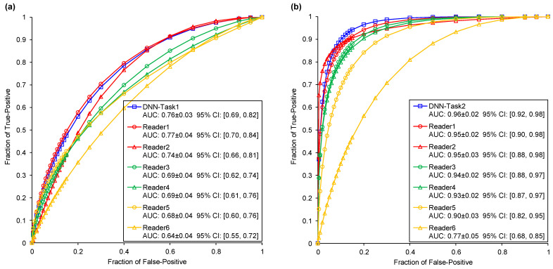
This study aims to develop a deep neural network (DNN)-based two-stage risk stratification model for early lung adenocarcinomas in CT images, and investigate the performance compared with practicing radiologists. A total of 2393 GGNs were retrospectively collected from 2105 patients in four centers. All the pathologic results of GGNs were obtained from surgically resected specimens. A two-stage deep neural network was developed based on the 3D residual network and atrous convolution module to diagnose benign and malignant GGNs (Task1) and classify between invasive adenocarcinoma (IA) and non-IA for these malignant GGNs (Task2). A multi-reader multi-case observer study with six board-certified radiologists' (average experience 11 years, range 2-28 years) participation was conducted to evaluate the model capability. DNN yielded area under the receiver operating characteristic curve (AUC) values of 0.76 ± 0.03 (95% confidence interval (CI): (0.69, 0.82)) and 0.96 ± 0.02 (95% CI: (0.92, 0.98)) for Task1 and Task2, which were equivalent to or higher than radiologists in the senior group with average AUC values of 0.76 and 0.95, respectively ( > 0.05). With the CT image slice thickness increasing from 1.15 mm ± 0.36 to 1.73 mm ± 0.64, DNN performance decreased 0.08 and 0.22 for the two tasks. The results demonstrated (1) a positive trend between the diagnostic performance and radiologist's experience, (2) the DNN yielded equivalent or even higher performance in comparison with senior radiologists, and (3) low image resolution decreased model performance in predicting the risks of GGNs. Once tested prospectively in clinical practice, the DNN could have the potential to assist doctors in precision diagnosis and treatment of early lung adenocarcinoma.
本研究旨在开发一种基于深度神经网络(DNN)的两阶段风险分层模型,用于CT图像中的早期肺腺癌,并与执业放射科医生比较其性能。回顾性收集了四个中心2105例患者的2393个磨玻璃结节(GGN)。所有GGN的病理结果均来自手术切除标本。基于3D残差网络和空洞卷积模块开发了一个两阶段深度神经网络,以诊断良性和恶性GGN(任务1),并对这些恶性GGN进行浸润性腺癌(IA)和非IA分类(任务2)。进行了一项多读者多病例观察研究,有六位获得委员会认证的放射科医生(平均经验11年,范围2 - 28年)参与,以评估模型能力。DNN在任务1和任务2中的受试者工作特征曲线下面积(AUC)值分别为0.76±0.03(95%置信区间(CI):(0.69,0.82))和0.96±0.02(95%CI:(0.92,0.98)),分别相当于或高于高级组放射科医生,其平均AUC值分别为0.76和0.95(P>0.05)。随着CT图像切片厚度从1.15 mm±0.36增加到1.73 mm±0.64,DNN在两项任务中的性能分别下降了0.08和0.22。结果表明:(1)诊断性能与放射科医生经验之间呈正相关趋势;(2)与高级放射科医生相比,DNN产生了相当甚至更高的性能;(3)低图像分辨率会降低模型预测GGN风险的性能。一旦在临床实践中进行前瞻性测试,DNN可能有潜力协助医生对早期肺腺癌进行精准诊断和治疗。