• 文献检索
  • 文档翻译
  • 深度研究
  • 学术资讯
  • Suppr Zotero 插件Zotero 插件
  • 邀请有礼
  • 套餐&价格
  • 历史记录
应用&插件
Suppr Zotero 插件Zotero 插件浏览器插件Mac 客户端Windows 客户端微信小程序
定价
高级版会员购买积分包购买API积分包
服务
文献检索文档翻译深度研究API 文档MCP 服务
关于我们
关于 Suppr公司介绍联系我们用户协议隐私条款
关注我们

Suppr 超能文献

核心技术专利:CN118964589B侵权必究
粤ICP备2023148730 号-1Suppr @ 2026

文献检索

告别复杂PubMed语法,用中文像聊天一样搜索,搜遍4000万医学文献。AI智能推荐,让科研检索更轻松。

立即免费搜索

文件翻译

保留排版,准确专业,支持PDF/Word/PPT等文件格式,支持 12+语言互译。

免费翻译文档

深度研究

AI帮你快速写综述,25分钟生成高质量综述,智能提取关键信息,辅助科研写作。

立即免费体验

用于肺炎分类的四种多类框架及其在使用七种深度学习人工智能模型的X射线扫描中的验证

Four Types of Multiclass Frameworks for Pneumonia Classification and Its Validation in X-ray Scans Using Seven Types of Deep Learning Artificial Intelligence Models.

作者信息

Jain Pankaj K, Sharma Neeraj, Kalra Mannudeep K, Viskovic Klaudija, Saba Luca, Suri Jasjit S

机构信息

School of Biomedical Engineering, Indian Institute of Technology (BHU), Varanasi 221005, India.

Department of Radiology, Massachusetts General Hospital, Boston, MA 02115, USA.

出版信息

Diagnostics (Basel). 2022 Mar 7;12(3):652. doi: 10.3390/diagnostics12030652.

DOI:10.3390/diagnostics12030652
PMID:35328205
原文链接:https://pmc.ncbi.nlm.nih.gov/articles/PMC8946935/
Abstract

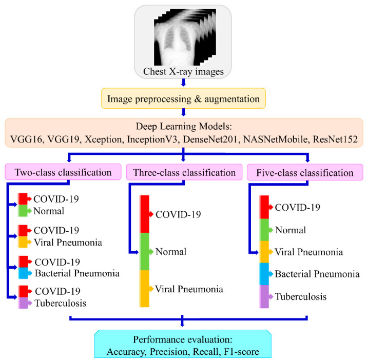
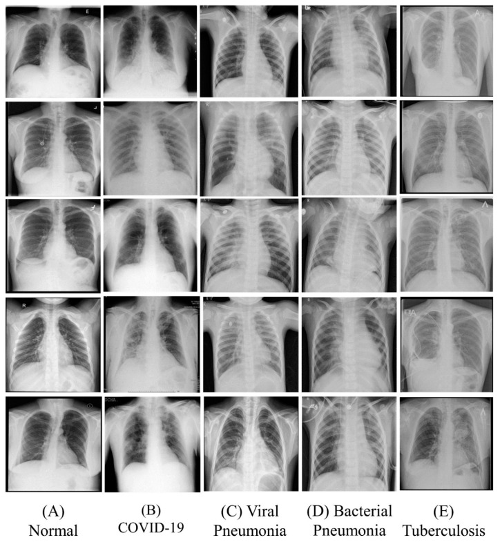
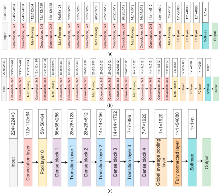
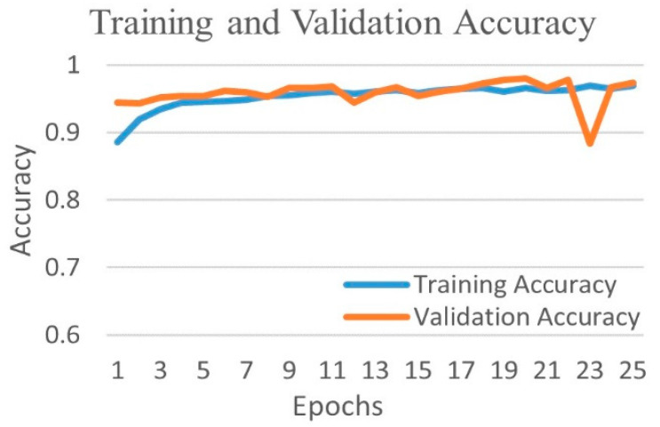
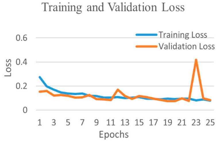
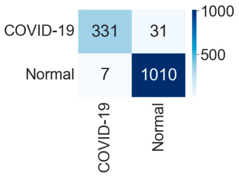
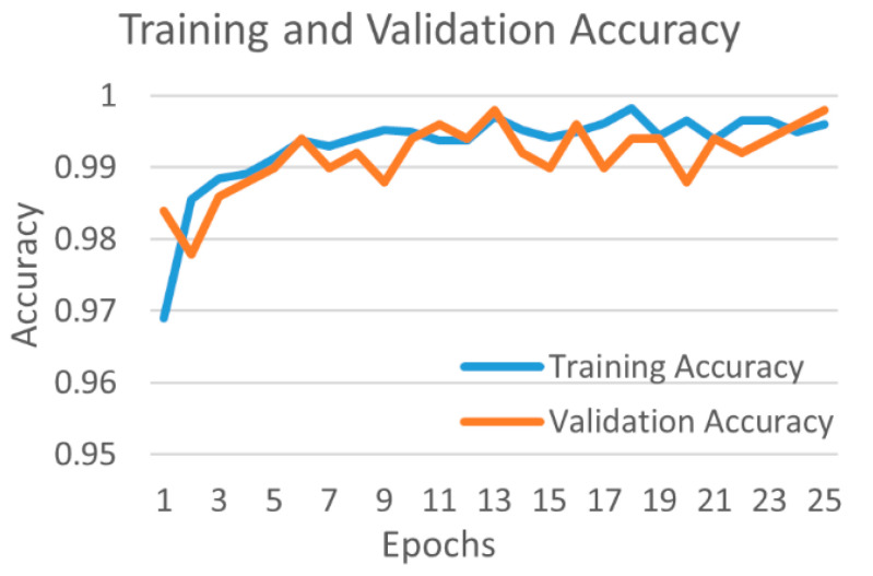
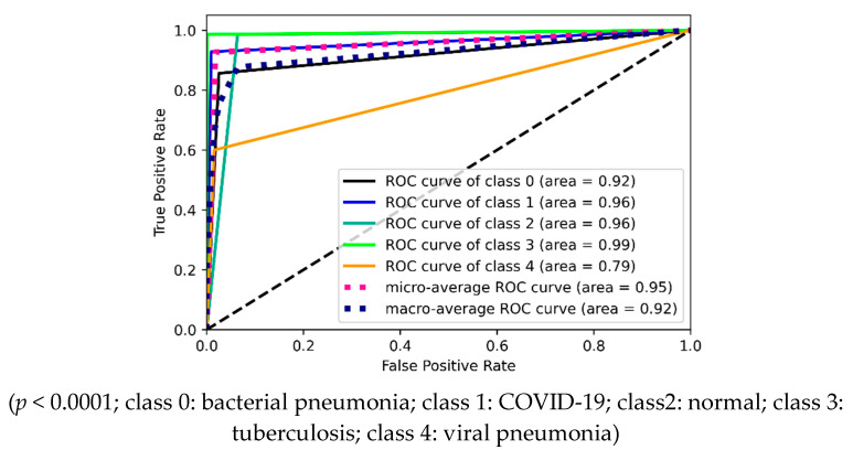
Background and Motivation: The novel coronavirus causing COVID-19 is exceptionally contagious, highly mutative, decimating human health and life, as well as the global economy, by consistent evolution of new pernicious variants and outbreaks. The reverse transcriptase polymerase chain reaction currently used for diagnosis has major limitations. Furthermore, the multiclass lung classification X-ray systems having viral, bacterial, and tubercular classes—including COVID-19—are not reliable. Thus, there is a need for a robust, fast, cost-effective, and easily available diagnostic method. Method: Artificial intelligence (AI) has been shown to revolutionize all walks of life, particularly medical imaging. This study proposes a deep learning AI-based automatic multiclass detection and classification of pneumonia from chest X-ray images that are readily available and highly cost-effective. The study has designed and applied seven highly efficient pre-trained convolutional neural networks—namely, VGG16, VGG19, DenseNet201, Xception, InceptionV3, NasnetMobile, and ResNet152—for classification of up to five classes of pneumonia. Results: The database consisted of 18,603 scans with two, three, and five classes. The best results were using DenseNet201, VGG16, and VGG16, respectively having accuracies of 99.84%, 96.7%, 92.67%; sensitivity of 99.84%, 96.63%, 92.70%; specificity of 99.84, 96.63%, 92.41%; and AUC of 1.0, 0.97, 0.92 (p < 0.0001 for all), respectively. Our system outperformed existing methods by 1.2% for the five-class model. The online system takes <1 s while demonstrating reliability and stability. Conclusions: Deep learning AI is a powerful paradigm for multiclass pneumonia classification.

摘要

背景与动机

引发新冠肺炎的新型冠状病毒具有极强的传染性和高度的变异性,通过不断演变出新的有害变种和疫情爆发,严重损害人类健康和生命以及全球经济。目前用于诊断的逆转录聚合酶链反应存在重大局限性。此外,包括新冠肺炎在内的具有病毒、细菌和结核类别分类的多类别肺部X射线系统并不可靠。因此,需要一种强大、快速、经济高效且易于获得的诊断方法。方法:人工智能(AI)已被证明能彻底改变各行各业,尤其是医学成像领域。本研究提出一种基于深度学习人工智能的方法,用于从胸部X射线图像中自动进行多类别肺炎检测和分类,这些图像易于获取且成本效益高。该研究设计并应用了七个高效的预训练卷积神经网络,即VGG16、VGG19、DenseNet201、Xception、InceptionV3、NasnetMobile和ResNet152,用于多达五类肺炎的分类。结果:数据库包含18603次扫描,分为两类、三类和五类。最佳结果分别使用DenseNet201、VGG16和VGG16,准确率分别为99.84%、96.7%、92.67%;灵敏度分别为99.84%、96.63%、92.70%;特异性分别为99.84、96.63%、92.41%;曲线下面积分别为1.0、0.97、0.92(所有p<0.0001)。对于五类模型,我们的系统比现有方法性能高出1.2%。在线系统在展示可靠性和稳定性的同时,处理时间不到1秒。结论:深度学习人工智能是多类别肺炎分类的强大范例。

https://cdn.ncbi.nlm.nih.gov/pmc/blobs/c85b/8946935/b89f488350b4/diagnostics-12-00652-g027.jpg
https://cdn.ncbi.nlm.nih.gov/pmc/blobs/c85b/8946935/d8874464bd40/diagnostics-12-00652-g001.jpg
https://cdn.ncbi.nlm.nih.gov/pmc/blobs/c85b/8946935/42bce309d48d/diagnostics-12-00652-g002.jpg
https://cdn.ncbi.nlm.nih.gov/pmc/blobs/c85b/8946935/476f3650ee5f/diagnostics-12-00652-g003a.jpg
https://cdn.ncbi.nlm.nih.gov/pmc/blobs/c85b/8946935/70e5acb7c5b2/diagnostics-12-00652-g004.jpg
https://cdn.ncbi.nlm.nih.gov/pmc/blobs/c85b/8946935/55d4211c2301/diagnostics-12-00652-g005.jpg
https://cdn.ncbi.nlm.nih.gov/pmc/blobs/c85b/8946935/a2e22d64939d/diagnostics-12-00652-g006.jpg
https://cdn.ncbi.nlm.nih.gov/pmc/blobs/c85b/8946935/cc3f2c835e41/diagnostics-12-00652-g007.jpg
https://cdn.ncbi.nlm.nih.gov/pmc/blobs/c85b/8946935/744cdeb8ad4d/diagnostics-12-00652-g008.jpg
https://cdn.ncbi.nlm.nih.gov/pmc/blobs/c85b/8946935/0bd3e8dbb7b6/diagnostics-12-00652-g009.jpg
https://cdn.ncbi.nlm.nih.gov/pmc/blobs/c85b/8946935/d40e3f933cc5/diagnostics-12-00652-g010.jpg
https://cdn.ncbi.nlm.nih.gov/pmc/blobs/c85b/8946935/08e18fd8ce3f/diagnostics-12-00652-g011.jpg
https://cdn.ncbi.nlm.nih.gov/pmc/blobs/c85b/8946935/65c39ebaab44/diagnostics-12-00652-g012.jpg
https://cdn.ncbi.nlm.nih.gov/pmc/blobs/c85b/8946935/63f00112ad79/diagnostics-12-00652-g013.jpg
https://cdn.ncbi.nlm.nih.gov/pmc/blobs/c85b/8946935/9049cbb70374/diagnostics-12-00652-g014.jpg
https://cdn.ncbi.nlm.nih.gov/pmc/blobs/c85b/8946935/bf696cc9bb4d/diagnostics-12-00652-g015.jpg
https://cdn.ncbi.nlm.nih.gov/pmc/blobs/c85b/8946935/7af2795d320a/diagnostics-12-00652-g016.jpg
https://cdn.ncbi.nlm.nih.gov/pmc/blobs/c85b/8946935/625901ded01f/diagnostics-12-00652-g017.jpg
https://cdn.ncbi.nlm.nih.gov/pmc/blobs/c85b/8946935/34992e34f070/diagnostics-12-00652-g018.jpg
https://cdn.ncbi.nlm.nih.gov/pmc/blobs/c85b/8946935/f0ad4e71b6e0/diagnostics-12-00652-g019.jpg
https://cdn.ncbi.nlm.nih.gov/pmc/blobs/c85b/8946935/13ff95cf51ce/diagnostics-12-00652-g020.jpg
https://cdn.ncbi.nlm.nih.gov/pmc/blobs/c85b/8946935/636d9dc78107/diagnostics-12-00652-g021.jpg
https://cdn.ncbi.nlm.nih.gov/pmc/blobs/c85b/8946935/b59294f63010/diagnostics-12-00652-g022.jpg
https://cdn.ncbi.nlm.nih.gov/pmc/blobs/c85b/8946935/0ca215c7d4d9/diagnostics-12-00652-g023.jpg
https://cdn.ncbi.nlm.nih.gov/pmc/blobs/c85b/8946935/35f3b74dab6c/diagnostics-12-00652-g024.jpg
https://cdn.ncbi.nlm.nih.gov/pmc/blobs/c85b/8946935/87a906963cbd/diagnostics-12-00652-g025.jpg
https://cdn.ncbi.nlm.nih.gov/pmc/blobs/c85b/8946935/1df5dbdfbb1e/diagnostics-12-00652-g026.jpg
https://cdn.ncbi.nlm.nih.gov/pmc/blobs/c85b/8946935/b89f488350b4/diagnostics-12-00652-g027.jpg

相似文献

1
Four Types of Multiclass Frameworks for Pneumonia Classification and Its Validation in X-ray Scans Using Seven Types of Deep Learning Artificial Intelligence Models.用于肺炎分类的四种多类框架及其在使用七种深度学习人工智能模型的X射线扫描中的验证
Diagnostics (Basel). 2022 Mar 7;12(3):652. doi: 10.3390/diagnostics12030652.
2
Segmentation-Based Classification Deep Learning Model Embedded with Explainable AI for COVID-19 Detection in Chest X-ray Scans.基于分割的分类深度学习模型,嵌入可解释人工智能用于胸部X光扫描中的新冠肺炎检测
Diagnostics (Basel). 2022 Sep 2;12(9):2132. doi: 10.3390/diagnostics12092132.
3
COV-RadNet: A Deep Convolutional Neural Network for Automatic Detection of COVID-19 from Chest X-Rays and CT Scans.COV-RadNet:一种用于从胸部X光和CT扫描中自动检测新冠病毒肺炎的深度卷积神经网络。
Comput Methods Programs Biomed Update. 2022;2:100064. doi: 10.1016/j.cmpbup.2022.100064. Epub 2022 Aug 25.
4
Detection of COVID-19 in X-ray Images Using Densely Connected Squeeze Convolutional Neural Network (DCSCNN): Focusing on Interpretability and Explainability of the Black Box Model.基于密集连接挤压卷积神经网络(DCSCNN)的 X 射线图像中 COVID-19 的检测:聚焦黑箱模型的可解释性和可说明性。
Sensors (Basel). 2022 Dec 18;22(24):9983. doi: 10.3390/s22249983.
5
Application of deep learning to identify COVID-19 infection in posteroanterior chest X-rays.深度学习在前后位胸部 X 光片中识别 COVID-19 感染的应用。
Clin Imaging. 2021 Dec;80:268-273. doi: 10.1016/j.clinimag.2021.07.004. Epub 2021 Jul 24.
6
Automatic coronavirus disease 2019 diagnosis based on chest radiography and deep learning - Success story or dataset bias?基于胸部 X 光和深度学习的新型冠状病毒病 2019 自动诊断——成功案例还是数据集偏差?
Med Phys. 2022 Feb;49(2):978-987. doi: 10.1002/mp.15419. Epub 2022 Jan 12.
7
PulDi-COVID: Chronic obstructive pulmonary (lung) diseases with COVID-19 classification using ensemble deep convolutional neural network from chest X-ray images to minimize severity and mortality rates.PulDi-COVID:利用胸部X光图像的集成深度卷积神经网络对患有COVID-19的慢性阻塞性肺疾病进行分类,以降低严重程度和死亡率。
Biomed Signal Process Control. 2023 Mar;81:104445. doi: 10.1016/j.bspc.2022.104445. Epub 2022 Nov 30.
8
A hybrid explainable ensemble transformer encoder for pneumonia identification from chest X-ray images.一种用于从胸部 X 光图像中识别肺炎的混合可解释集成式变压器编码器。
J Adv Res. 2023 Jun;48:191-211. doi: 10.1016/j.jare.2022.08.021. Epub 2022 Sep 7.
9
Multiclass magnetic resonance imaging brain tumor classification using artificial intelligence paradigm.使用人工智能范式的多类别磁共振成像脑肿瘤分类
Comput Biol Med. 2020 Jul;122:103804. doi: 10.1016/j.compbiomed.2020.103804. Epub 2020 May 30.
10
A preliminary analysis of AI based smartphone application for diagnosis of COVID-19 using chest X-ray images.基于人工智能的智能手机应用程序利用胸部X光图像诊断新冠肺炎的初步分析。
Expert Syst Appl. 2021 Nov 30;183:115401. doi: 10.1016/j.eswa.2021.115401. Epub 2021 Jun 12.

引用本文的文献

1
An Artificial Intelligence-Based Non-Invasive Approach for Cardiovascular Disease Risk Stratification in Obstructive Sleep Apnea Patients: A Narrative Review.一种基于人工智能的阻塞性睡眠呼吸暂停患者心血管疾病风险分层的非侵入性方法:叙述性综述。
Rev Cardiovasc Med. 2024 Dec 28;25(12):463. doi: 10.31083/j.rcm2512463. eCollection 2024 Dec.
2
Confidence-Aware Severity Assessment of Lung Disease from Chest X-Rays Using Deep Neural Network on a Multi-Reader Dataset.基于多阅片者数据集利用深度神经网络对胸部X光片进行肺部疾病的置信度感知严重程度评估
J Imaging Inform Med. 2025 Apr;38(2):793-803. doi: 10.1007/s10278-024-01151-5. Epub 2024 Aug 20.
3

本文引用的文献

1
Learning discriminative representation for image classification.学习用于图像分类的判别性表示。
Knowl Based Syst. 2021 Dec 5;233. doi: 10.1016/j.knosys.2021.107517. Epub 2021 Sep 23.
2
An artificial intelligence framework and its bias for brain tumor segmentation: A narrative review.一种用于脑肿瘤分割的人工智能框架及其偏差:一篇综述。
Comput Biol Med. 2022 Apr;143:105273. doi: 10.1016/j.compbiomed.2022.105273. Epub 2022 Feb 19.
3
The COVID-19 Infection Diffusion in the US and Japan: A Graph-Theoretical Approach.新冠病毒在美国和日本的感染扩散:一种基于图论的方法。
UltraAIGenomics: Artificial Intelligence-Based Cardiovascular Disease Risk Assessment by Fusion of Ultrasound-Based Radiomics and Genomics Features for Preventive, Personalized and Precision Medicine: A Narrative Review.
超人工智能基因组学:基于超声影像组学和基因组学特征融合的人工智能心血管疾病风险评估用于预防、个性化和精准医学:一篇综述
Rev Cardiovasc Med. 2024 May 22;25(5):184. doi: 10.31083/j.rcm2505184. eCollection 2024 May.
4
Four Transformer-Based Deep Learning Classifiers Embedded with an Attention U-Net-Based Lung Segmenter and Layer-Wise Relevance Propagation-Based Heatmaps for COVID-19 X-ray Scans.四种基于Transformer的深度学习分类器,嵌入了基于注意力U-Net的肺部分割器和基于逐层相关性传播的热图,用于COVID-19 X光扫描。
Diagnostics (Basel). 2024 Jul 16;14(14):1534. doi: 10.3390/diagnostics14141534.
5
Teleradiology and technology innovations in radiology: status in India and its role in increasing access to primary health care.远程放射学与放射学技术创新:印度现状及其在增加初级卫生保健可及性方面的作用。
Lancet Reg Health Southeast Asia. 2023 Apr 14;23:100195. doi: 10.1016/j.lansea.2023.100195. eCollection 2024 Apr.
6
Ensemble Deep Learning Derived from Transfer Learning for Classification of COVID-19 Patients on Hybrid Deep-Learning-Based Lung Segmentation: A Data Augmentation and Balancing Framework.基于迁移学习的集成深度学习用于基于混合深度学习的肺部分割对新冠肺炎患者进行分类:一种数据增强与平衡框架
Diagnostics (Basel). 2023 Jun 2;13(11):1954. doi: 10.3390/diagnostics13111954.
7
Role of Ensemble Deep Learning for Brain Tumor Classification in Multiple Magnetic Resonance Imaging Sequence Data.集成深度学习在多磁共振成像序列数据脑肿瘤分类中的作用
Diagnostics (Basel). 2023 Jan 28;13(3):481. doi: 10.3390/diagnostics13030481.
8
Cardiovascular/Stroke Risk Stratification in Diabetic Foot Infection Patients Using Deep Learning-Based Artificial Intelligence: An Investigative Study.使用基于深度学习的人工智能对糖尿病足感染患者进行心血管/中风风险分层:一项调查研究。
J Clin Med. 2022 Nov 19;11(22):6844. doi: 10.3390/jcm11226844.
9
Segmentation-Based Classification Deep Learning Model Embedded with Explainable AI for COVID-19 Detection in Chest X-ray Scans.基于分割的分类深度学习模型,嵌入可解释人工智能用于胸部X光扫描中的新冠肺炎检测
Diagnostics (Basel). 2022 Sep 2;12(9):2132. doi: 10.3390/diagnostics12092132.
10
COVID-19 Classification from Chest X-Ray Images: A Framework of Deep Explainable Artificial Intelligence.基于深度可解释人工智能的 COVID-19 胸片分类框架。
Comput Intell Neurosci. 2022 Jul 14;2022:4254631. doi: 10.1155/2022/4254631. eCollection 2022.
Biology (Basel). 2022 Jan 13;11(1):125. doi: 10.3390/biology11010125.
4
Cardiovascular disease detection using machine learning and carotid/femoral arterial imaging frameworks in rheumatoid arthritis patients.利用机器学习和颈动脉/股动脉成像框架在类风湿关节炎患者中进行心血管疾病检测。
Rheumatol Int. 2022 Feb;42(2):215-239. doi: 10.1007/s00296-021-05062-4. Epub 2022 Jan 11.
5
Unseen Artificial Intelligence-Deep Learning Paradigm for Segmentation of Low Atherosclerotic Plaque in Carotid Ultrasound: A Multicenter Cardiovascular Study.用于颈动脉超声中低动脉粥样硬化斑块分割的不可见人工智能深度学习范式:一项多中心心血管研究。
Diagnostics (Basel). 2021 Dec 2;11(12):2257. doi: 10.3390/diagnostics11122257.
6
Ten Fast Transfer Learning Models for Carotid Ultrasound Plaque Tissue Characterization in Augmentation Framework Embedded with Heatmaps for Stroke Risk Stratification.用于在嵌入热图的增强框架中进行颈动脉超声斑块组织特征分析以实现中风风险分层的十种快速迁移学习模型
Diagnostics (Basel). 2021 Nov 15;11(11):2109. doi: 10.3390/diagnostics11112109.
7
The Chaotic Behavior of the Spread of Infection During the COVID-19 Pandemic in the United States and Globally.美国及全球新冠疫情期间感染传播的混沌行为
IEEE Access. 2021 Jun 2;9:80692-80702. doi: 10.1109/ACCESS.2021.3085240. eCollection 2021.
8
Deep learning based detection and analysis of COVID-19 on chest X-ray images.基于深度学习的胸部X光图像中新型冠状病毒肺炎的检测与分析
Appl Intell (Dordr). 2021;51(3):1690-1700. doi: 10.1007/s10489-020-01902-1. Epub 2020 Oct 9.
9
Thrombotic sinusoiditis and local diffuse intrasinusoidal coagulation in the liver of subjects affected by COVID-19: the evidence from histology and scanning electron microscopy.COVID-19 感染者肝脏中的血栓性窦状隙炎和局部弥漫性窦内弥散性凝血:组织学和扫描电子显微镜证据。
Eur Rev Med Pharmacol Sci. 2021 Oct;25(19):5904-5912. doi: 10.26355/eurrev_202110_26866.
10
COVID-19 diagnosis from chest x-rays: developing a simple, fast, and accurate neural network.基于胸部X光片的COVID-19诊断:开发一种简单、快速且准确的神经网络。
Health Inf Sci Syst. 2021 Oct 12;9(1):36. doi: 10.1007/s13755-021-00166-4. eCollection 2021 Dec.