Palmer D A, Douglas J T
University of Hawaii, Honolulu 96822.
J Clin Microbiol. 1989 Oct;27(10):2331-7. doi: 10.1128/jcm.27.10.2331-2337.1989.
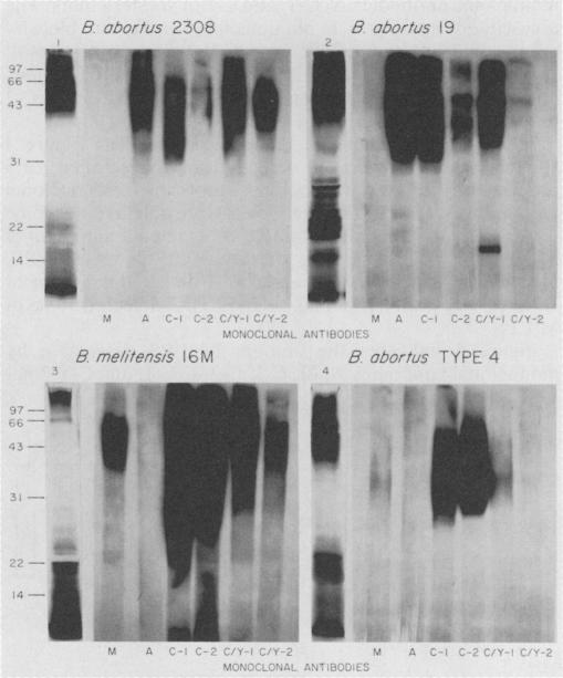
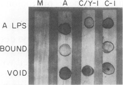
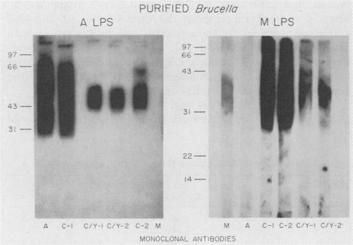
Monoclonal antibodies which bind Brucella A lipopolysaccharide (LPS)-specific, M LPS-specific, or cross-reactive epitopes were used as reagents in quantitative dot blot, Western blot (immunoblot), and immunoprecipitation analysis of Brucella whole cells, whole-cell extracts, and purified LPS preparations. This set of monoclonal antibodies detected four unique epitopes on Brucella LPS. The specificity of monoclonal antibodies reactive with Brucella unique (A and M) and common (C and C/Y) LPS epitopes was demonstrated by blot analysis. The serotype specificity of monoclonal antibodies for A LPS of B. abortus 1119.3 or M LPS of Brucella melitensis 16M was confirmed. Type C monoclonal antibodies recognized epitopes on Brucella A and M LPS and did not cross-react with Yersinia enterocolitica O:9. In Western blots, type C monoclonal antibodies were bound by epitopes on Brucella A and M LPSs ranging in Mrs from 30,000 to 70,000, relative to marker proteins. Type C/Y monoclonal antibodies were cross-reactive with Y. enterocolitica O:9 and recognized Brucella A LPS epitopes with a restricted Mr ranging only from 40,000 to 50,000, relative to marker proteins. Type C/Y monoclonal antibodies also displayed a more restricted pattern of binding to Brucella M LPS. The monoclonal antibodies were able to detect 5 to 50 pg of a purified A LPS preparation in dot blots. The limits of detection by the monoclonal antibodies of a purified M LPS preparation ranged from 0.05 to 50 pg. Monoclonal antibody analysis of whole-cell preparations also demonstrated quantitative differences in the presence of the respective epitopes. The binding profiles of the monoclonal antibodies to Brucella whole cells varied between acetone- and chloroform-killed organisms as well as between species and strains. The lower limit of detection of any whole-cell preparation by the dot blot technique was 10(5) CFU. Binding profiles in Western blots and endotoxin activity of immunoprecipitates obtained with these monoclonal antibodies further defined the Brucella LPS antigens. These monoclonal antibodies and the techniques described may be useful in monitoring the antigenic content of Brucella vaccines and diagnostics.
与布鲁氏菌 A 型脂多糖(LPS)特异性、M 型 LPS 特异性或交叉反应性表位结合的单克隆抗体,被用作试剂,用于对布鲁氏菌全细胞、全细胞提取物和纯化的 LPS 制剂进行定量斑点印迹、蛋白质免疫印迹(免疫印迹)和免疫沉淀分析。这组单克隆抗体在布鲁氏菌 LPS 上检测到四个独特的表位。通过印迹分析证明了与布鲁氏菌独特(A 和 M)和常见(C 和 C/Y)LPS 表位反应的单克隆抗体的特异性。确认了单克隆抗体对流产布鲁氏菌 1119.3 的 A LPS 或羊种布鲁氏菌 16M 的 M LPS 的血清型特异性。C 型单克隆抗体识别布鲁氏菌 A 和 M LPS 上的表位,且不与小肠结肠炎耶尔森菌 O:9 发生交叉反应。在蛋白质免疫印迹中,相对于标记蛋白,C 型单克隆抗体与分子量在 30,000 至 70,000 之间的布鲁氏菌 A 和 M LPS 上的表位结合。C/Y 型单克隆抗体与小肠结肠炎耶尔森菌 O:9 发生交叉反应,且相对于标记蛋白,识别分子量仅在 40,000 至 50,000 之间的布鲁氏菌 A LPS 表位。C/Y 型单克隆抗体与布鲁氏菌 M LPS 的结合模式也更具局限性。这些单克隆抗体在斑点印迹中能够检测到 5 至 50 pg 的纯化 A LPS 制剂。单克隆抗体对纯化 M LPS 制剂的检测限在 0.05 至 50 pg 之间。对全细胞制剂的单克隆抗体分析也显示了各自表位存在的定量差异。单克隆抗体与布鲁氏菌全细胞的结合谱在丙酮和氯仿灭活的生物体之间以及不同种和菌株之间有所不同。斑点印迹技术对任何全细胞制剂的检测下限为 10(5) CFU。用这些单克隆抗体获得的蛋白质免疫印迹中的结合谱和免疫沉淀物的内毒素活性进一步明确了布鲁氏菌 LPS 抗原。这些单克隆抗体和所描述的技术可能有助于监测布鲁氏菌疫苗和诊断试剂的抗原含量。