Department of Radiology and Biomedical Imaging, University of California San Francisco, San Francisco, CA, USA.
Department of Radiology and Biomedical Imaging, University of California San Francisco, San Francisco, CA, USA; UCSF/UC Berkeley Graduate Group in Bioengineering, USA.
Neuroimage Clin. 2018 Aug 4;20:498-505. doi: 10.1016/j.nicl.2018.08.002. eCollection 2018.
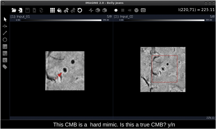
With extensive research efforts in place to address the clinical relevance of cerebral microbleeds (CMBs), there remains a need for fast and accurate methods to detect and quantify CMB burden. Although some computer-aided detection algorithms have been proposed in the literature with high sensitivity, their specificity remains consistently poor. More sophisticated machine learning methods appear to be promising in their ability to minimize false positives (FP) through high-level feature extraction and the discrimination of hard-mimics. To achieve superior performance, these methods require sizable amounts of precisely labelled training data. Here we present a user-guided tool for semi-automated CMB detection and volume segmentation, offering high specificity for routine use and FP labelling capabilities to ease and expedite the process of generating labelled training data.
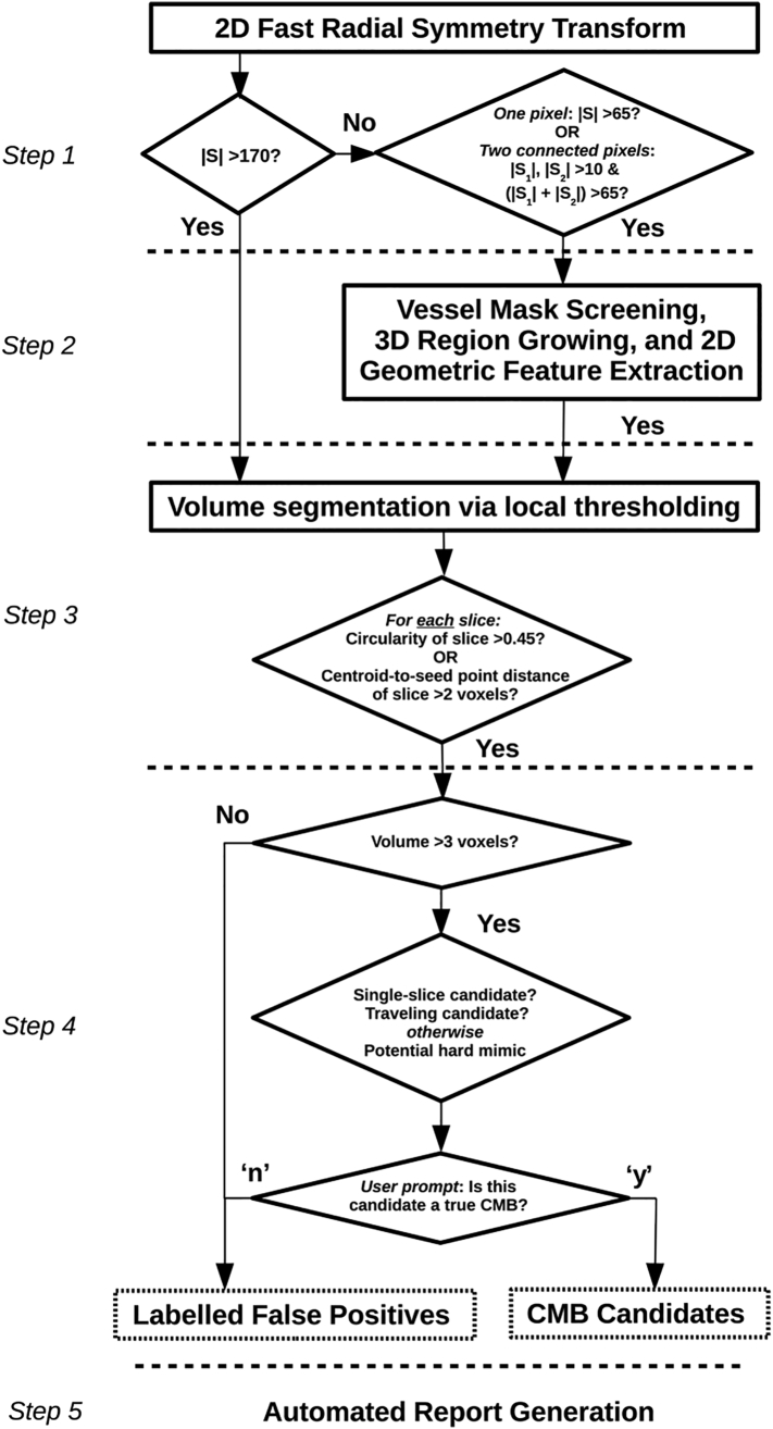
Existing computer-aided detection methods reported by our group were extended to include fully-automated segmentation and user-guided CMB classification with FP labelling. The algorithm's performance was evaluated on a test set of ten patients exhibiting radiotherapy-induced CMBs on MR images.
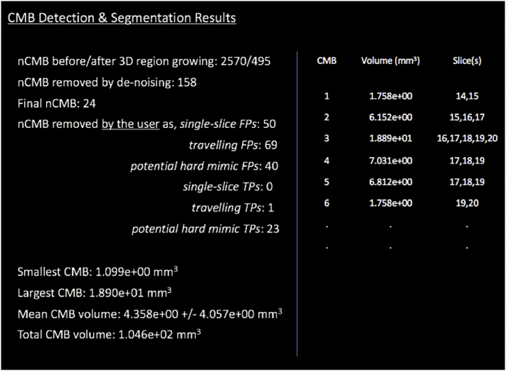
The initial algorithm's base sensitivity was maintained at 86.7%. FP's were reduced to inter-rater variations and segmentation results were in 98% agreement with ground truth labelling. There was an approximate 5-fold reduction in the time users spent evaluating CMB burden with the algorithm versus without computer aid. The Intra-class Correlation Coefficient for inter-rater agreement was 0.97 CI[0.92,0.99].
This development serves as a valuable tool for routine evaluation of CMB burden and data labelling to improve CMB classification with machine learning. The algorithm is available to the public on GitHub (https://github.com/LupoLab-UCSF/CMB_labeler).
尽管已经进行了广泛的研究来解决脑微出血(CMB)的临床相关性问题,但仍需要快速准确的方法来检测和量化 CMB 负担。尽管文献中已经提出了一些具有高灵敏度的计算机辅助检测算法,但它们的特异性仍然很差。更复杂的机器学习方法似乎在通过高级特征提取和硬模仿的区分来最小化假阳性(FP)方面具有很大的潜力。为了获得卓越的性能,这些方法需要大量精确标记的训练数据。在这里,我们提出了一种用于半自动 CMB 检测和体积分割的用户引导工具,该工具具有高特异性,可用于常规使用,并且具有 FP 标记功能,可简化和加快生成标记训练数据的过程。
我们扩展了现有计算机辅助检测方法,包括全自动分割和用户引导的 CMB 分类和 FP 标记。该算法在十名患有放射治疗诱导的 CMB 的患者的测试集上进行了评估。
初始算法的基本灵敏度保持在 86.7%。FP 减少到了观察者间的变异,分割结果与地面实况标记的一致性达到 98%。与没有计算机辅助的情况下相比,用户评估 CMB 负担的时间减少了约 5 倍。观察者间一致性的组内相关系数为 0.97 CI[0.92,0.99]。
该开发为常规评估 CMB 负担和数据标记提供了有价值的工具,以改善机器学习的 CMB 分类。该算法可在 GitHub 上(https://github.com/LupoLab-UCSF/CMB_labeler)获得。