Universidad Peruana Cayetano Heredia, Laboratorio de Malaria, Laboratiorios de Investigacion y Dessarrollo, Facultad de Ciencias y Filosofia, Av. Honorio Delgado 430 SMP, Lima, Peru.
Intellectual Ventures, 3150 139 AVE SE, Bellevue, WA, 98005, USA.
Malar J. 2018 Sep 25;17(1):339. doi: 10.1186/s12936-018-2493-0.
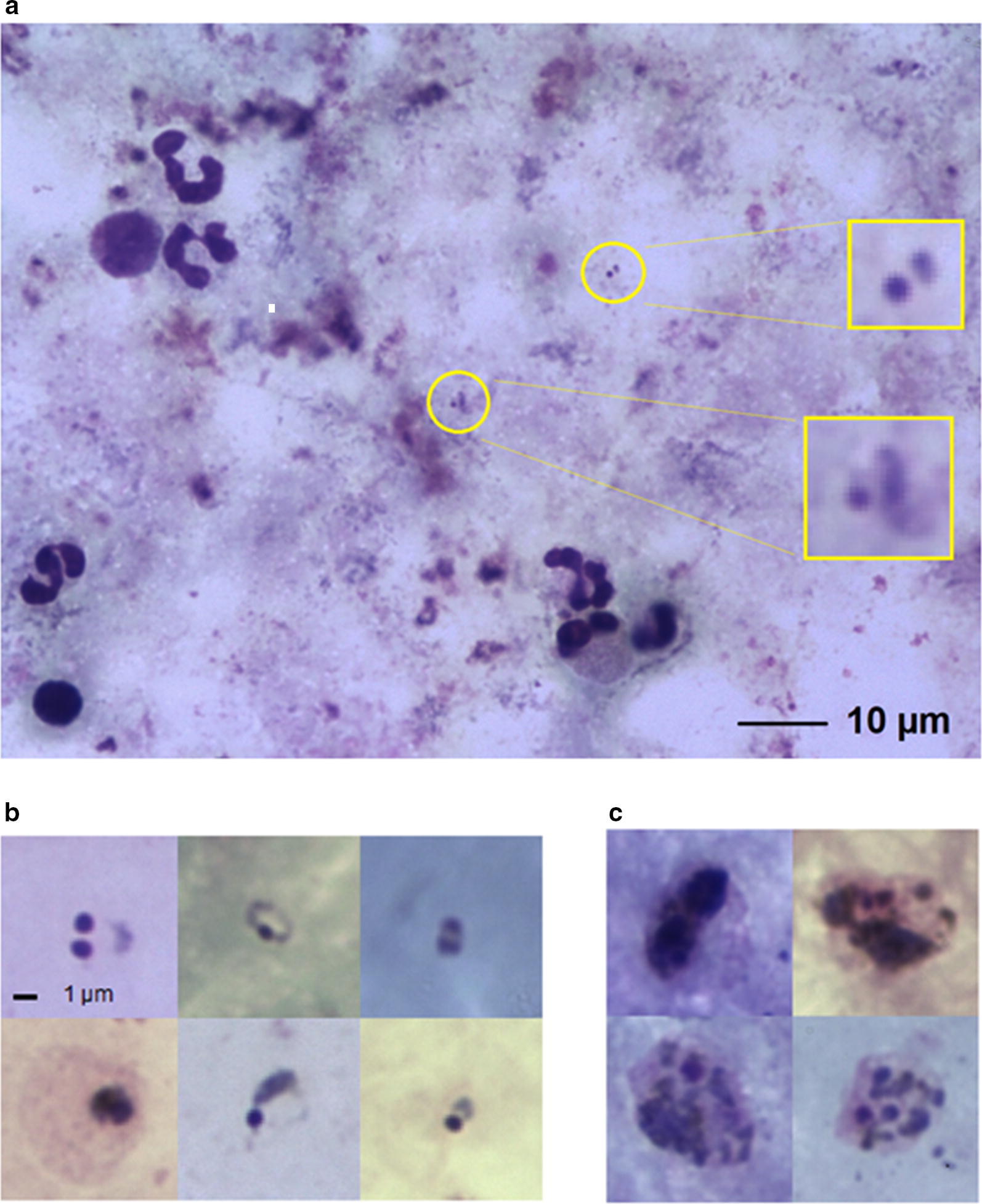
Microscopic examination of Giemsa-stained blood films remains a major form of diagnosis in malaria case management, and is a reference standard for research. However, as with other visualization-based diagnoses, accuracy depends on individual technician performance, making standardization difficult and reliability poor. Automated image recognition based on machine-learning, utilizing convolutional neural networks, offers potential to overcome these drawbacks. A prototype digital microscope device employing an algorithm based on machine-learning, the Autoscope, was assessed for its potential in malaria microscopy. Autoscope was tested in the Iquitos region of Peru in 2016 at two peripheral health facilities, with routine microscopy and PCR as reference standards. The main outcome measures include sensitivity and specificity of diagnosis of malaria from Giemsa-stained blood films, using PCR as reference.
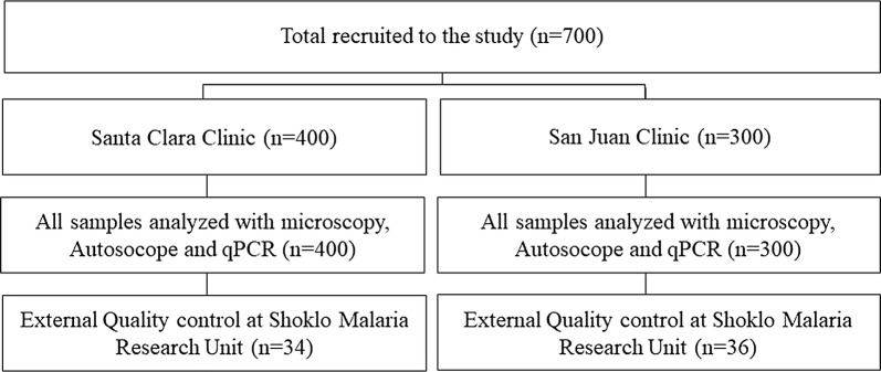
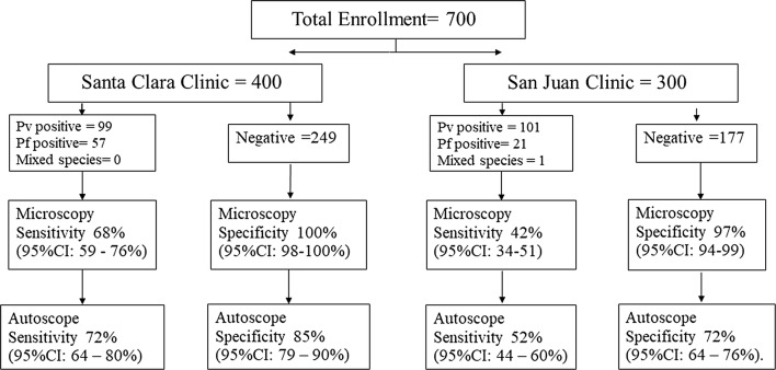
A cross-sectional, observational trial was conducted at two peripheral primary health facilities in Peru. 700 participants were enrolled with the criteria: (1) age between 5 and 75 years, (2) history of fever in the last 3 days or elevated temperature on admission, (3) informed consent. The main outcome measures included sensitivity and specificity of diagnosis of malaria from Giemsa-stained blood films, using PCR as reference.
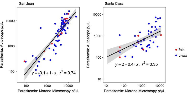
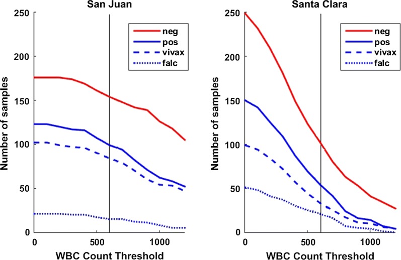
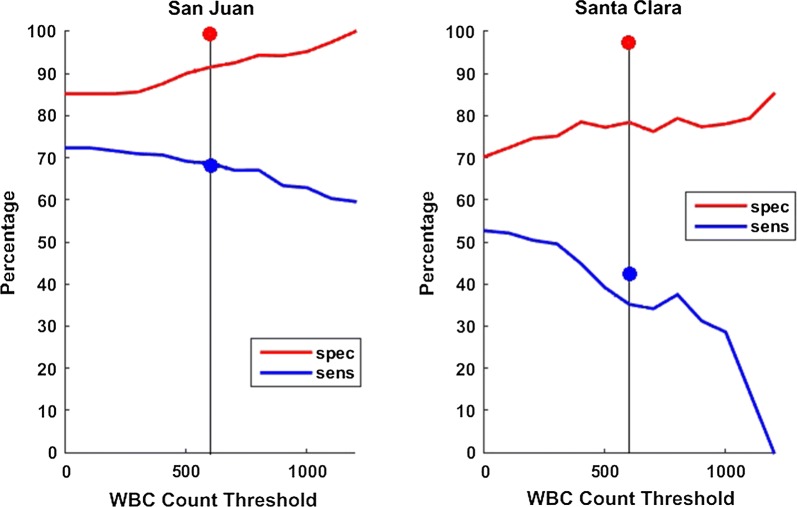
At the San Juan clinic, sensitivity of Autoscope for diagnosing malaria was 72% (95% CI 64-80%), and specificity was 85% (95% CI 79-90%). Microscopy performance was similar to Autoscope, with sensitivity 68% (95% CI 59-76%) and specificity 100% (95% CI 98-100%). At San Juan, 85% of prepared slides had a minimum of 600 WBCs imaged, thus meeting Autoscope's design assumptions. At the second clinic, Santa Clara, the sensitivity of Autoscope was 52% (95% CI 44-60%) and specificity was 70% (95% CI 64-76%). Microscopy performance at Santa Clara was 42% (95% CI 34-51) and specificity was 97% (95% CI 94-99). Only 39% of slides from Santa Clara met Autoscope's design assumptions regarding WBCs imaged.
Autoscope's diagnostic performance was on par with routine microscopy when slides had adequate blood volume to meet its design assumptions, as represented by results from the San Juan clinic. Autoscope's diagnostic performance was poorer than routine microscopy on slides from the Santa Clara clinic, which generated slides with lower blood volumes. Results of the study reflect both the potential for artificial intelligence to perform tasks currently conducted by highly-trained experts, and the challenges of replicating the adaptiveness of human thought processes.
吉姆萨染色血片的显微镜检查仍然是疟疾病例管理中的主要诊断形式,也是研究的参考标准。然而,与其他基于可视化的诊断一样,准确性取决于个体技术员的表现,因此难以标准化,可靠性也较差。基于机器学习的自动图像识别技术,利用卷积神经网络,为克服这些缺点提供了潜力。一种名为 Autoscope 的基于机器学习算法的原型数字显微镜设备,用于评估其在疟疾显微镜检查中的潜力。Autoscope 于 2016 年在秘鲁伊基托斯地区的两家基层医疗机构进行了测试,以常规显微镜检查和 PCR 作为参考标准。主要的观察指标包括使用 PCR 作为参考时,从吉姆萨染色血片中诊断疟疾的敏感性和特异性。
在秘鲁的两家基层初级医疗机构进行了一项横断面、观察性试验。符合以下标准的 700 名参与者被纳入研究:(1)年龄在 5 至 75 岁之间;(2)过去 3 天有发热史或入院时体温升高;(3)知情同意。主要的观察指标包括使用 PCR 作为参考时,从吉姆萨染色血片中诊断疟疾的敏感性和特异性。
在圣胡安诊所,Autoscope 诊断疟疾的敏感性为 72%(95%CI 64-80%),特异性为 85%(95%CI 79-90%)。显微镜检查的性能与 Autoscope 相似,敏感性为 68%(95%CI 59-76%),特异性为 100%(95%CI 98-100%)。在圣胡安,85%的准备好的载玻片上至少有 600 个白细胞成像,因此符合 Autoscope 的设计假设。在第二家诊所,圣克拉拉,Autoscope 的敏感性为 52%(95%CI 44-60%),特异性为 70%(95%CI 64-76%)。圣克拉拉的显微镜检查结果为 42%(95%CI 34-51%),特异性为 97%(95%CI 94-99%)。只有 39%的圣克拉拉载玻片符合 Autoscope 关于白细胞成像的设计假设。
当载玻片具有足够的血量以满足其设计假设时,Autoscope 的诊断性能与常规显微镜检查相当,这反映了圣胡安诊所的结果。Autoscope 在圣克拉拉诊所的载玻片上的诊断性能比常规显微镜检查差,因为圣克拉拉诊所的载玻片产生的血量较低。研究结果反映了人工智能执行目前由高技能专家执行的任务的潜力,以及复制人类思维过程适应性的挑战。