Das Anupam
Royal Global University, Guwahati, Assam 781033 India.
Multimed Tools Appl. 2022;81(4):5407-5441. doi: 10.1007/s11042-021-11787-y. Epub 2021 Dec 22.
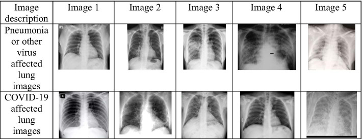
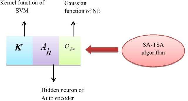
COVID-19 disease is a major health calamity in twentieth century, in which the infection is spreading at the global level. Developing countries like Bangladesh, India, and others are still facing a delay in recognizing COVID-19 cases. Hence, there is a need for immediate recognition with perfect identification of infection. This clear visualization helps to save the life of suspected COVID-19 patients. With the help of traditional RT-PCR testing, the combination of medical images and deep learning classifiers delivers more hopeful results with high accuracy in the prediction and recognition of COVID-19 cases. COVID-19 disease is recently researched through sample chest X-ray images, which have already proven its efficiency in lung diseases. To emphasize corona virus testing methods and to control the community spreading, the automatic detection process of COVID-19 is processed through the detailed medication reports from medical images. Although there are numerous challenges in the manual understanding of traces in COVID-19 infection from X-ray, the subtle differences among normal and infected X-rays can be traced by the data patterns of Convolutional Neural Network (CNN). To improve the detection performance of CNN, this paper plans to develop an Ensemble Learning with CNN-based Deep Features (EL-CNN-DF). In the initial phase, image scaling and median filtering perform the pre-processing of the chest X-ray images gathered from the benchmark source. The second phase is lung segmentation, which is the significant step for COVID detection. It is accomplished by the Adaptive Activation Function-based U-Net (AAF-U-Net). Once the lungs are segmented, it is subjected to novel EL-CNN-DF, in which the deep features are extracted from the pooling layer of CNN, and the fully connected layer of CNN are replaced with the three classifiers termed "Support Vector Machine (SVM), Autoencoder, Naive Bayes (NB)". The final detection of COVID-19 is done by these classifiers, in which high ranking strategy is utilized. As a modification, a Self Adaptive-Tunicate Swarm Algorithm (SA-TSA) is adopted as a boosting algorithm to enhance the performance of segmentation and detection. The overall analysis has shown that the precision of the enhanced CNN by using SA-TSA was 1.02%, 4.63%, 3.38%, 1.62%, 1.51% and 1.04% better than SVM, autoencoder, NB, Ensemble, RNN and LSTM respectively. The comparative performance analysis on existing model proves that the proposed algorithm is better than other algorithms in terms of segmentation and classification of COVID-19 detection.
新冠病毒病是20世纪的一场重大健康灾难,其感染正在全球范围内蔓延。像孟加拉国、印度等发展中国家仍在新冠病毒病病例的识别上存在延迟。因此,需要立即进行识别并准确鉴定感染情况。这种清晰的可视化有助于挽救疑似新冠病毒病患者的生命。借助传统的逆转录聚合酶链反应(RT-PCR)检测,医学图像与深度学习分类器相结合,在新冠病毒病病例的预测和识别方面能以高精度给出更有希望的结果。近期通过胸部X光样本图像对新冠病毒病进行了研究,胸部X光样本图像在肺部疾病方面已证明了其有效性。为了强调冠状病毒检测方法并控制社区传播,新冠病毒病的自动检测过程通过医学图像的详细用药报告来进行。尽管从X光手动理解新冠病毒感染痕迹存在诸多挑战,但卷积神经网络(CNN)的数据模式可以追踪正常X光和感染X光之间的细微差异。为了提高CNN的检测性能,本文计划开发一种基于CNN深度特征的集成学习(EL-CNN-DF)。在初始阶段,图像缩放和中值滤波对从基准源收集的胸部X光图像进行预处理。第二阶段是肺部分割,这是新冠病毒检测的重要步骤。它由基于自适应激活函数的U型网络(AAF-U-Net)完成。一旦肺部被分割,就对其应用新颖的EL-CNN-DF,其中深度特征从CNN的池化层提取,并且CNN的全连接层被替换为三个分类器,即“支持向量机(SVM)、自动编码器、朴素贝叶斯(NB)”。新冠病毒病的最终检测由这些分类器完成,其中采用了高排名策略。作为一种改进,采用自适应-樽海鞘群算法(SA-TSA)作为增强算法来提高分割和检测性能。总体分析表明,使用SA-TSA增强后的CNN在精度上分别比SVM、自动编码器、NB、集成、循环神经网络(RNN)和长短期记忆网络(LSTM)高1.02%、4.63%、3.38%、1.62%、1.51%和1.04%。对现有模型的比较性能分析证明,所提出的算法在新冠病毒病检测的分割和分类方面优于其他算法。