Guduru R, Liang P, Yousef M, Horstmyer J, Khizroev S
1Center for Personalized Nanomedicine, Florida International University, 11200 SW 8th ST, Miami, Florida 33199 USA.
2Department of Electrical and Computer Engineering, Florida International University, Miami, Florida 33174 USA.
Bioelectron Med. 2018 Aug 6;4:10. doi: 10.1186/s42234-018-0012-9. eCollection 2018.
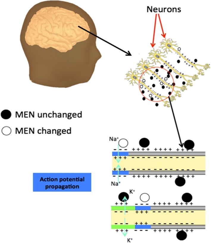
Neurodegenerative diseases are devastating diagnoses. Examining local electric fields in response to neural activity in real time could shed light on understanding the origins of these diseases. To date, there has not been found a way to directly map these fields without interfering with the electric circuitry of the brain. This theoretical study is focused on a nanotechnology concept to overcome the challenge of brain electric field mapping in real time. The paper shows that coupling the magnetoelectric effect of multiferroic nanoparticles, known as magnetoelectric nanoparticles (MENs), with the ultra-fast and high-sensitivity imaging capability of the recently emerged magnetic particle imaging (MPI) can enable wirelessly conducted electric-field mapping with specifications to meet the requirements for monitoring neural activity in real time.
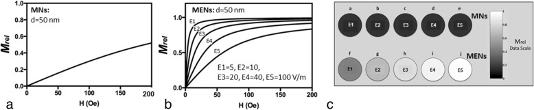
The MPI signal is numerically simulated on a realistic human brain template obtained from BrainWeb, while brain segmentation was performed with BrainSuite software. The finite element mesh is generated with Computer Geometry Algorithm Library. The effect of MENs is modeled through local point magnetization changes according to the magnetoelectric effect.
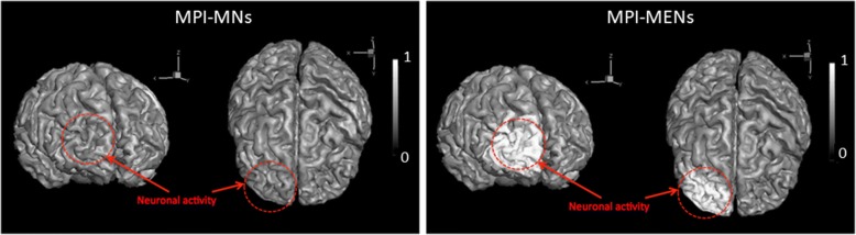
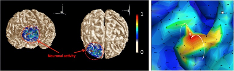
It is shown that, unlike traditional magnetic nanoparticles, MENs, when coupled with MPI, provide information containing electric field's spatial and temporal patterns due to local neural activity with signal sensitivities adequate for detection of minute changes at the sub-cellular level corresponding to early stage disease processes.
Like no other nanoparticles known to date, MENs coupled with MPI can be used for mapping electric field activity of the brain at the sub-neuronal level in real time. The potential applications span from prevention and treatment of neurodegenerative diseases to paving the way to fundamental understanding and reverse engineering the brain.
神经退行性疾病是极具毁灭性的诊断结果。实时检测响应神经活动的局部电场有助于深入了解这些疾病的起源。迄今为止,尚未找到一种在不干扰大脑电路的情况下直接绘制这些电场的方法。这项理论研究聚焦于一种纳米技术概念,以克服实时进行脑电场映射的挑战。本文表明,将多铁性纳米颗粒(称为磁电纳米颗粒,MENs)的磁电效应与最近出现的磁粒子成像(MPI)的超快速和高灵敏度成像能力相结合,能够实现无线进行电场映射,其规格满足实时监测神经活动的要求。
在从BrainWeb获得的真实人脑模板上对MPI信号进行数值模拟,同时使用BrainSuite软件进行脑部分割。使用计算机几何算法库生成有限元网格。根据磁电效应,通过局部点磁化变化对MENs的作用进行建模。
结果表明,与传统磁性纳米颗粒不同,MENs与MPI结合时,由于局部神经活动提供包含电场空间和时间模式的信息,其信号灵敏度足以检测对应于疾病早期过程的亚细胞水平的微小变化。
与迄今所知的其他纳米颗粒不同,MENs与MPI结合可用于实时绘制亚神经元水平的脑电场活动。其潜在应用范围从神经退行性疾病的预防和治疗到为深入理解大脑和大脑逆向工程铺平道路。