College of Computer and Information Engineering, Tianjin Normal University, Tianjin, China.
Department of Computer Science and Engineering, Fairfield University, Fairfield, USA.
Comput Intell Neurosci. 2020 Mar 30;2020:8975078. doi: 10.1155/2020/8975078. eCollection 2020.
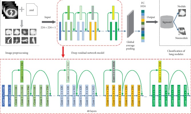
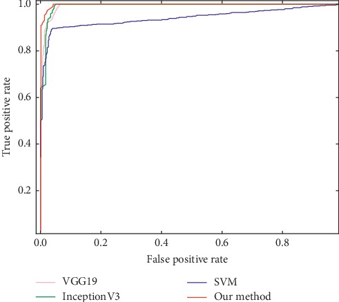
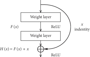
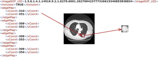
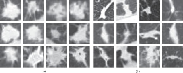
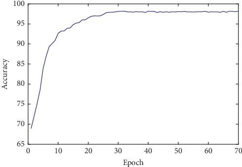
The classification process of lung nodule detection in a traditional computer-aided detection (CAD) system is complex, and the classification result is heavily dependent on the performance of each step in lung nodule detection, causing low classification accuracy and high false positive rate. In order to alleviate these issues, a lung nodule classification method based on a deep residual network is proposed. Abandoning traditional image processing methods and taking the 50-layer ResNet network structure as the initial model, the deep residual network is constructed by combining residual learning and migration learning. The proposed approach is verified by conducting experiments on the lung computed tomography (CT) images from the publicly available LIDC-IDRI database. An average accuracy of 98.23% and a false positive rate of 1.65% are obtained based on the ten-fold cross-validation method. Compared with the conventional support vector machine (SVM)-based CAD system, the accuracy of our method improved by 9.96% and the false positive rate decreased by 6.95%, while the accuracy improved by 1.75% and 2.42%, respectively, and the false positive rate decreased by 2.07% and 2.22%, respectively, in contrast to the VGG19 model and InceptionV3 convolutional neural networks. The experimental results demonstrate the effectiveness of our proposed method in lung nodule classification for CT images.
传统计算机辅助检测(CAD)系统中的肺结节检测分类过程复杂,分类结果严重依赖于肺结节检测中每个步骤的性能,导致分类精度低,假阳性率高。为了缓解这些问题,提出了一种基于深度残差网络的肺结节分类方法。该方法放弃了传统的图像处理方法,以 50 层 ResNet 网络结构作为初始模型,通过结合残差学习和迁移学习构建深度残差网络。该方法在公开的 LIDC-IDRI 数据库的肺部 CT 图像上进行了验证。基于十折交叉验证方法,获得了平均准确率为 98.23%和假阳性率为 1.65%。与传统的基于支持向量机(SVM)的 CAD 系统相比,我们的方法的准确率提高了 9.96%,假阳性率降低了 6.95%,而与 VGG19 模型和 InceptionV3 卷积神经网络相比,准确率分别提高了 1.75%和 2.42%,假阳性率分别降低了 2.07%和 2.22%。实验结果表明,该方法在 CT 图像的肺结节分类中是有效的。