Collaborative Innovation Center for Biomedical Engineering, Wuhan National Laboratory for Optoelectronics-Huazhong University of Science and Technology, Wuhan, Hubei, China.
Britton Chance Center and MOE Key Laboratory for Biomedical Photonics, School of Engineering Sciences, Huazhong University of Science and Technology, Wuhan, Hubei, China.
Nat Commun. 2021 Sep 24;12(1):5639. doi: 10.1038/s41467-021-25296-x.
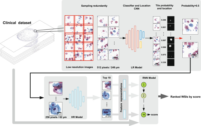
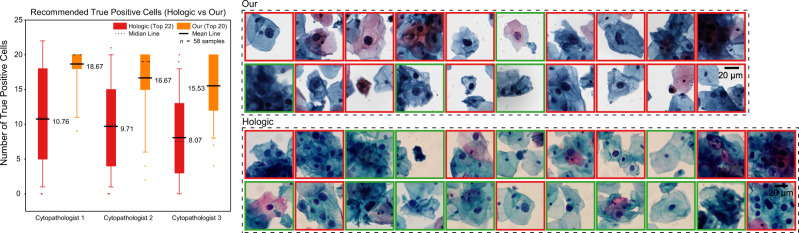
Computer-assisted diagnosis is key for scaling up cervical cancer screening. However, current recognition algorithms perform poorly on whole slide image (WSI) analysis, fail to generalize for diverse staining and imaging, and show sub-optimal clinical-level verification. Here, we develop a progressive lesion cell recognition method combining low- and high-resolution WSIs to recommend lesion cells and a recurrent neural network-based WSI classification model to evaluate the lesion degree of WSIs. We train and validate our WSI analysis system on 3,545 patient-wise WSIs with 79,911 annotations from multiple hospitals and several imaging instruments. On multi-center independent test sets of 1,170 patient-wise WSIs, we achieve 93.5% Specificity and 95.1% Sensitivity for classifying slides, comparing favourably to the average performance of three independent cytopathologists, and obtain 88.5% true positive rate for highlighting the top 10 lesion cells on 447 positive slides. After deployment, our system recognizes a one giga-pixel WSI in about 1.5 min.
计算机辅助诊断是扩大宫颈癌筛查的关键。然而,目前的识别算法在全切片图像(WSI)分析方面表现不佳,无法针对不同的染色和成像进行泛化,并且在临床水平的验证上表现不佳。在这里,我们开发了一种结合低分辨率和高分辨率 WSI 的渐进式病变细胞识别方法,用于推荐病变细胞,以及一种基于递归神经网络的 WSI 分类模型,用于评估 WSI 的病变程度。我们在来自多家医院和多种成像仪器的 3545 份患者 WSI 上进行了训练和验证,并进行了 79911 次注释。在来自 1170 份患者 WSI 的多中心独立测试集中,我们对幻灯片进行分类的特异性为 93.5%,敏感性为 95.1%,优于三位独立细胞病理学家的平均表现,并在 447 张阳性幻灯片上突出显示前 10 名病变细胞的真阳性率为 88.5%。部署后,我们的系统可以在大约 1.5 分钟内识别一个十亿像素的 WSI。